The CLCN1 Antibody (CAB5739) is a high-quality antibody developed for reliable detection and analysis of target proteins. This antibody, raised in rabbits, is highly specific to human samples and is validated for use in various applications, including Western blotting.CLCN1 plays a crucial role in maintaining muscle membrane excitability and proper muscle function. Dysregulation of CLCN1 has been implicated in muscle disorders such as myotonia congenita, making it a target of interest for researchers studying neuromuscular diseases.
This antibody is validated for use in WB, IHC-P, ELISA applications and has demonstrated reactivity against Human, Mouse, Rat samples.
Product Name:
CLCN1 Antibody
SKU:
CAB5739
Size:
20μL, 100μL
Reactivity:
Human, Mouse, Rat
Conjugate:
Unconjugated
Immunogen:
Recombinant protein (or fragment).This information is considered to be commercially sensitive.
Recommended starting concentration is 1 μg/mL. Please optimize the concentration based on your specific assay requirements.
Synonyms:
CLC1, CLCN1
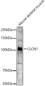
Positive Sample:
Mouse skeletal muscle
Cellular Localization:
Membrane, Multi-Pass Membrane Protein.
Calculated MW:
109kDa
Observed MW:
109kDa
The CLCN family of voltage-dependent chloride channel genes comprises nine members (CLCN1-7, Ka and Kb) which demonstrate quite diverse functional characteristics while sharing significant sequence homology. The protein encoded by this gene regulates the electric excitability of the skeletal muscle membrane. Mutations in this gene cause two forms of inherited human muscle disorders: recessive generalized myotonia congenita (Becker) and dominant myotonia (Thomsen). Alternative splicing results in multiple transcript variants.
Purification Method
Affinity purification
Gene ID
1180
RRID
AB_2766495
Buffer Information
Store at -20℃. Avoid freeze / thaw cycles. Buffer: PBS containing 50% glycerol, preserved with proclin300 or sodium azide, pH 7.3.
Western blot analysis of lysates from Mouse skeletal muscle, using CLCN1 Rabbit pAb (CAB5739) at 1:1000 dilution. Secondary antibody: HRP-conjugated Goat anti-Rabbit IgG (H+L) (CABS014) at 1:10000 dilution. Lysates/proteins: 25μg per lane. Blocking buffer: 3% nonfat dry milk in TBST. Detection: ECL Enhanced Kit (AbGn00021). Exposure time: 180s.
Immunohistochemistry analysis of paraffin-embedded Mouse skeletal muscle using CLCN1 Rabbit pAb (CAB5739) at dilution of 1:200 (40x lens). High pressure antigen retrieval performed with 0.01M Citrate buffer (pH 6.0) prior to IHC staining.