The DRG1 Antibody (CAB3987) is a high-quality antibody developed for reliable detection and analysis of target proteins. This antibody, produced in rabbits, demonstrates high reactivity with human samples and has been validated for use in Western blot applications.DRG1, also known as developmentally regulated GTP-binding protein 1, plays a crucial role in regulating cell growth and survival, making it a potential target for cancer research and drug development. By binding to the DRG1 protein, this antibody enables researchers to detect and analyze its expression in various cell types, providing important insights into its function and potential therapeutic applications.
This antibody is validated for use in WB, ELISA applications and has demonstrated reactivity against Human, Mouse, Rat samples.
Product Name:
DRG1 Antibody
SKU:
CAB3987
Size:
20μL, 100μL
Reactivity:
Human, Mouse, Rat
Conjugate:
Unconjugated
Immunogen:
Recombinant protein (or fragment).This information is considered to be commercially sensitive.
Recommended starting concentration is 1 μg/mL. Please optimize the concentration based on your specific assay requirements.
Synonyms:
NEDD3, DRG1
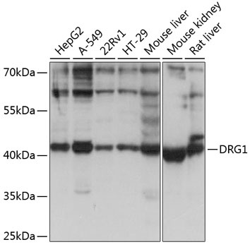
Positive Sample:
HepG2, A-549, 22Rv1, HT-29, Mouse liver, Mouse kidney, Rat liver
Cellular Localization:
Cytoplasm.
Calculated MW:
41kDa
Observed MW:
41kDa
Enables several functions, including GTPase activity; identical protein binding activity; and potassium ion binding activity. Involved in positive regulation of microtubule polymerization and regulation of mitotic spindle assembly. Located in cytosol and nuclear body. Part of polysome.
Purification Method
Affinity purification
Gene ID
4733
RRID
AB_2765432
Buffer Information
Store at -20℃. Avoid freeze / thaw cycles. Buffer: PBS with 0.01% thimerosal,50% glycerol,pH7.3.
Western blot analysis of various lysates using DRG1 Rabbit pAb (CAB3987) at 1:1000 dilution. Secondary antibody: HRP-conjugated Goat anti-Rabbit IgG (H+L) (CABS014) at 1:10000 dilution. Lysates/proteins: 25μg per lane. Blocking buffer: 3% nonfat dry milk in TBST. Detection: ECL Basic Kit (AbGn00020). Exposure time: 5s.