The DYRK2 Polyclonal Antibody (CAB24467) is a high-quality antibody developed for reliable detection and analysis of target proteins. This antibody, produced in rabbits, exhibits high reactivity with human tissue samples and has been validated for use in Western blot applications. By binding to the DYRK2 protein, researchers can effectively detect and analyze DYRK2 expression in a wide range of cell types, making it a valuable tool for studies in cancer and cell biology research.DYRK2 is known for its involvement in the regulation of key cellular processes such as cell division and programmed cell death, making it a promising target for investigating diseases like cancer and neurodegenerative disorders.
This antibody is validated for use in WB, IHC-P, ELISA applications and has demonstrated reactivity against Human, Rat samples.
Product Name:
DYRK2 Polyclonal Antibody
SKU:
CAB24467
Size:
20μL, 100μL
Reactivity:
Human, Rat
Conjugate:
Unconjugated
Immunogen:
Synthetic peptide. This information is considered to be commercially sensitive.
DYRK2 belongs to a family of protein kinases whose members are presumed to be involved in cellular growth and/or development. The family is defined by structural similarity of their kinase domains and their capability to autophosphorylate on tyrosine residues. DYRK2 has demonstrated tyrosine autophosphorylation and catalyzed phosphorylation of histones H3 and H2B in vitro. Two isoforms of DYRK2 have been isolated. The predominant isoform, isoform 1, lacks a 5' terminal insert.
Purification Method
Affinity purification
Gene ID
8445
Buffer Information
Store at -20℃. Avoid freeze / thaw cycles. Buffer: PBS containing 50% glycerol, preserved with proclin300 or sodium azide, pH 7.3.
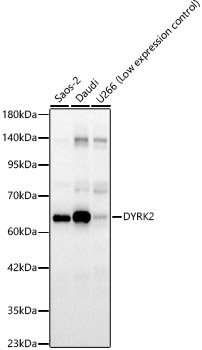
Western blot analysis of various lysates, using DYRK2 Rabbit pAb (CAB24467) at 1:700 dilution. Secondary antibody: HRP-conjugated Goat anti-Rabbit IgG (H+L) (CABS014) at 1:10000 dilution. Lysates/proteins: 25μg per lane. Blocking buffer: 3% nonfat dry milk in TBST. Detection: ECL Basic Kit (AbGn00020). Exposure time: 60s.
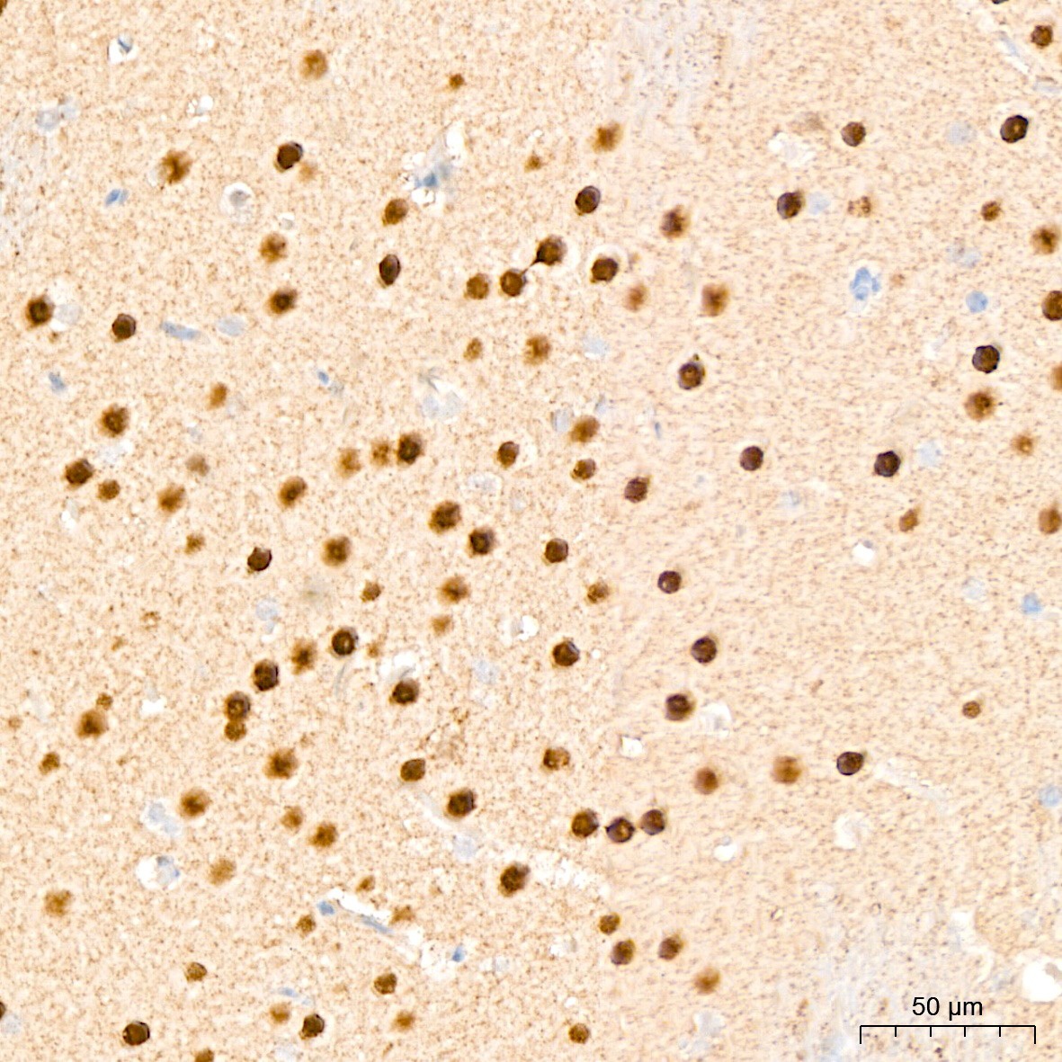
Immunohistochemistry analysis of paraffin-embedded Rat brain tissue using DYRK2 Rabbit pAb (CAB24467) at a dilution of 1:200 (40x lens). High pressure antigen retrieval was performed with 0.01M Citrate Buffer (pH 6.0) prior to IHC staining.
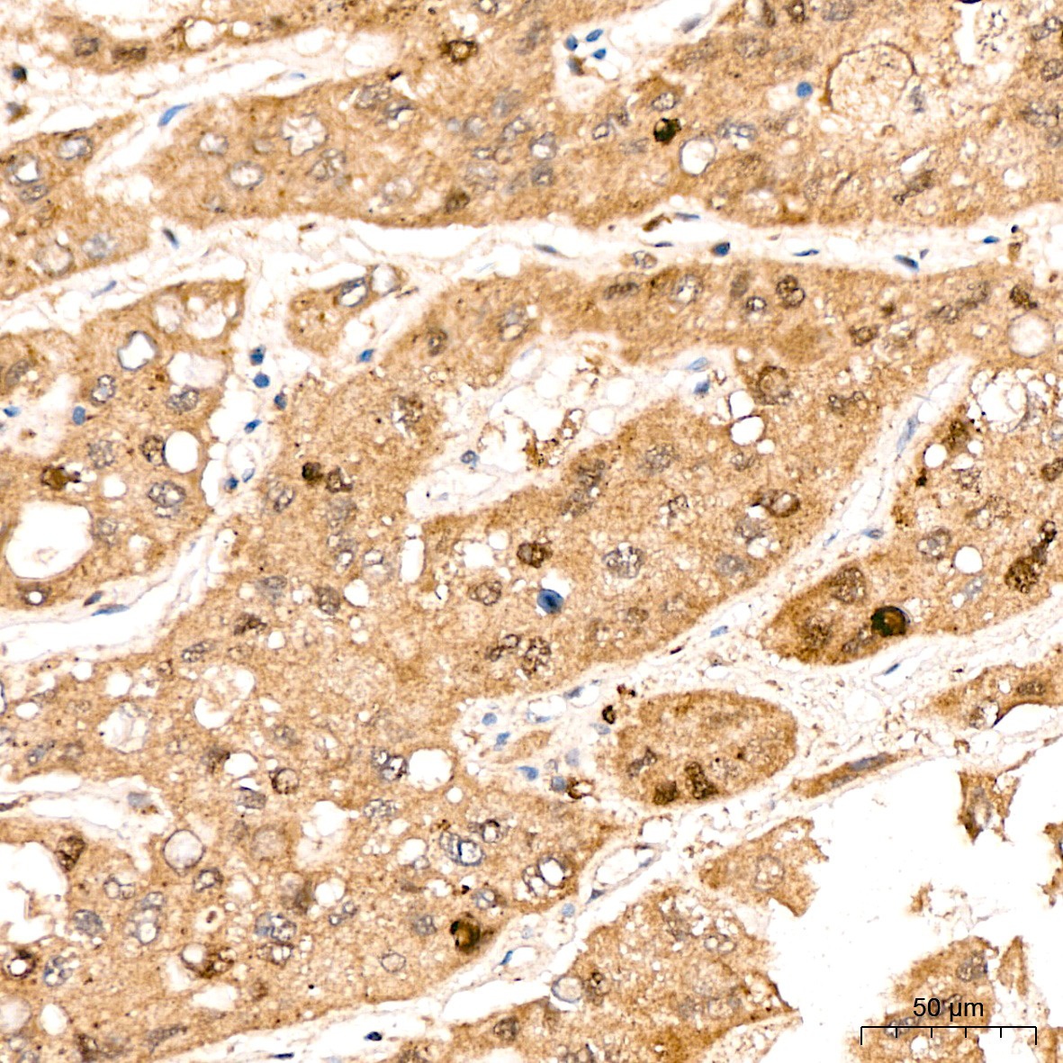
Immunohistochemistry analysis of paraffin-embedded Human liver cancer tissue using DYRK2 Rabbit pAb (CAB24467) at a dilution of 1:200 (40x lens). High pressure antigen retrieval was performed with 0.01M Citrate Buffer (pH 6.0) prior to IHC staining.
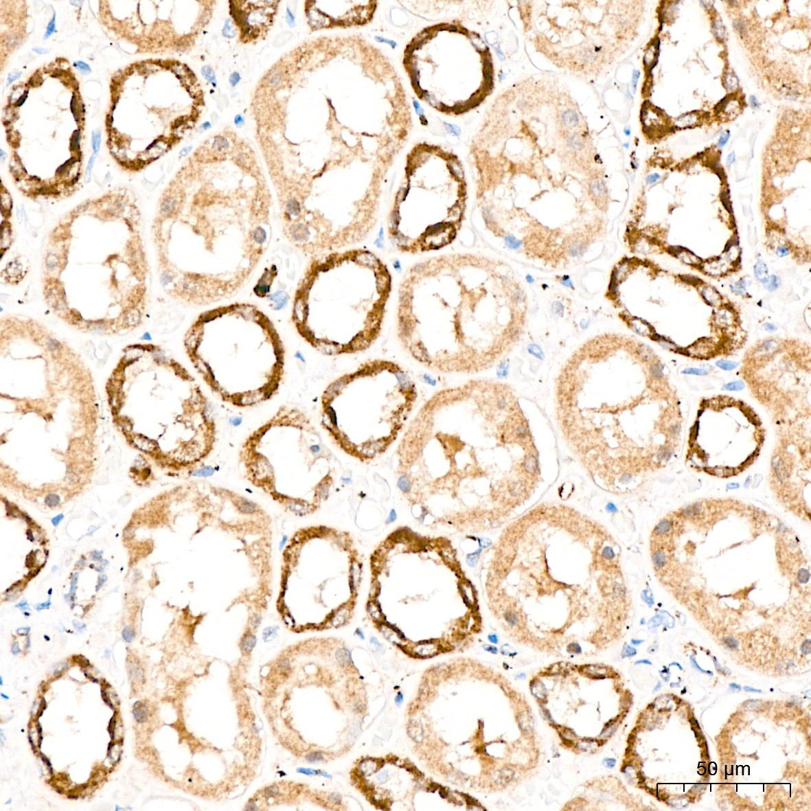
Immunohistochemistry analysis of paraffin-embedded Human kidney tissue using DYRK2 Rabbit pAb (CAB24467) at a dilution of 1:200 (40x lens). High pressure antigen retrieval was performed with 0.01M Citrate Buffer (pH 6.0) prior to IHC staining.
 at 1:700 dilution. Secondary antibody: HRP Goat Anti-Rabbit IgG (H+L) at 1:10000 dilution. Lysates/proteins: 25ug per lane. Blocking buffer: 3% nonfat dry milk in TBST.")
 at 1:700 dilution. Secondary antibody: HRP Goat Anti-Rabbit IgG (H+L) at 1:10000 dilution. Lysates/proteins: 25ug per lane. Blocking buffer: 3% nonfat dry milk in TBST.")


")