The eEF1A1 Antibody (CAB17857) is a high-quality antibody developed for reliable detection and analysis of target proteins. This antibody, produced in rabbits, is highly specific for human samples and has been validated for use in Western blot applications. By binding to the EEF1A1 protein, this antibody allows for precise detection and analysis in a variety of cell types, making it an ideal choice for studies in molecular biology and cancer research.EEF1A1 is a versatile protein involved in the elongation phase of protein synthesis and has been linked to various cellular processes, including cell proliferation, apoptosis, and cytoskeletal organization.
This antibody is validated for use in WB, IHC-P, IF/ICC, ELISA applications and has demonstrated reactivity against Human, Mouse, Rat samples.
Product Name:
eEF1A1 Antibody
SKU:
CAB17857
Size:
20μL, 100μL
Reactivity:
Human, Mouse, Rat
Conjugate:
Unconjugated
Immunogen:
Synthetic peptide. This information is considered to be commercially sensitive.
This gene encodes an isoform of the alpha subunit of the elongation factor-1 complex, which is responsible for the enzymatic delivery of aminoacyl tRNAs to the ribosome. This isoform (alpha 1) is expressed in brain, placenta, lung, liver, kidney, and pancreas, and the other isoform (alpha 2) is expressed in brain, heart and skeletal muscle. This isoform is identified as an autoantigen in 66% of patients with Felty syndrome. This gene has been found to have multiple copies on many chromosomes, some of which, if not all, represent different pseudogenes.
Purification Method
Affinity purification
Gene ID
1915
RRID
AB_2769265
Buffer Information
Store at -20℃. Avoid freeze / thaw cycles. Buffer: PBS containing 50% glycerol, preserved with proclin300 or sodium azide, pH 7.3.
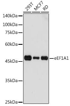
Western blot analysis of various lysates using eEF1A1 Rabbit pAb (CAB17857) at 1:1000 dilution. Secondary antibody: HRP-conjugated Goat anti-Rabbit IgG (H+L) (CABS014) at 1:10000 dilution. Lysates/proteins: 25μg per lane. Blocking buffer: 3% nonfat dry milk in TBST. Detection: ECL Basic Kit (AbGn00020). Exposure time: 3s.
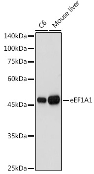
Western blot analysis of various lysates using eEF1A1 Rabbit pAb (CAB17857) at 1:1000 dilution. Secondary antibody: HRP-conjugated Goat anti-Rabbit IgG (H+L) (CABS014) at 1:10000 dilution. Lysates/proteins: 25μg per lane. Blocking buffer: 3% nonfat dry milk in TBST. Detection: ECL Basic Kit (AbGn00020). Exposure time: 10s.
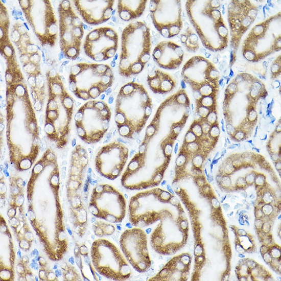
Immunohistochemistry analysis of paraffin-embedded Mouse kidney using eEF1A1 Rabbit pAb (CAB17857) at dilution of 1:100 (40x lens). High pressure antigen retrieval performed with 0.01M Citrate buffer (pH 6.0) prior to IHC staining.
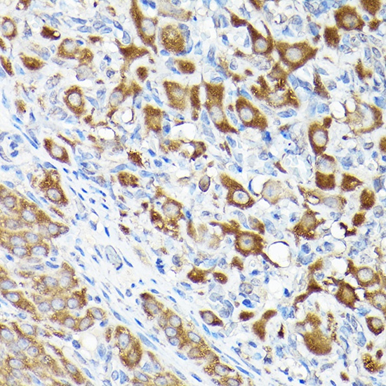
Immunohistochemistry analysis of paraffin-embedded Rat ovary using eEF1A1 Rabbit pAb (CAB17857) at dilution of 1:100 (40x lens). High pressure antigen retrieval performed with 0.01M Citrate buffer (pH 6.0) prior to IHC staining.
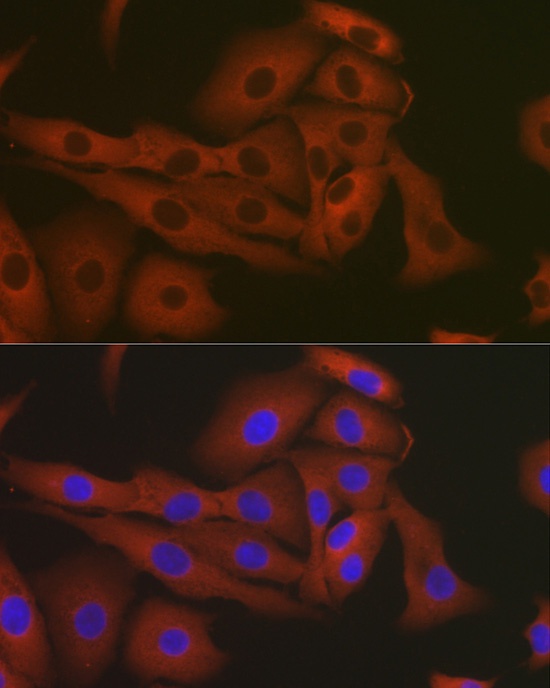
Immunofluorescence analysis of A-549 cells using eEF1A1 Rabbit pAb (CAB17857) at dilution of 1:50 (40x lens). Secondary antibody: Cy3-conjugated Goat anti-Rabbit IgG (H+L) (CABS007) at 1:500 dilution. Blue: DAPI for nuclear staining.
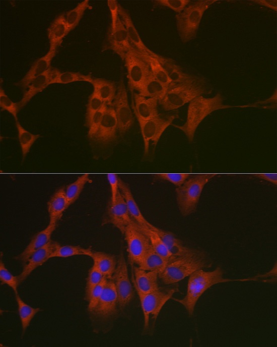
Immunofluorescence analysis of C6 cells using eEF1A1 Rabbit pAb (CAB17857) at dilution of 1:50 (40x lens). Secondary antibody: Cy3-conjugated Goat anti-Rabbit IgG (H+L) (CABS007) at 1:500 dilution. Blue: DAPI for nuclear staining.
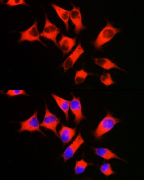
Immunofluorescence analysis of HeLa cells using eEF1A1 Rabbit pAb (CAB17857) at dilution of 1:50 (40x lens). Secondary antibody: Cy3-conjugated Goat anti-Rabbit IgG (H+L) (CABS007) at 1:500 dilution. Blue: DAPI for nuclear staining.
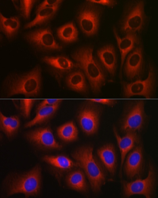
Immunofluorescence analysis of U2OS cells using eEF1A1 Rabbit pAb (CAB17857) at dilution of 1:50 (40x lens). Secondary antibody: Cy3-conjugated Goat anti-Rabbit IgG (H+L) (CABS007) at 1:500 dilution. Blue: DAPI for nuclear staining.