The FABP5 Antibody (CAB6373) is a high-quality antibody developed for reliable detection and analysis of target proteins. This antibody, generated in rabbits, exhibits high specificity and sensitivity towards FABP5 in human samples, making it ideal for use in Western blot applications.FABP5 is involved in the transport and metabolism of fatty acids, playing a crucial role in regulating lipid homeostasis and inflammatory responses. Dysregulation of FABP5 has been linked to various disease states, including metabolic disorders and inflammatory diseases.
This antibody is validated for use in WB, IHC-P, IF/ICC, ELISA applications and has demonstrated reactivity against Human, Mouse, Rat samples.
Product Name:
FABP5 Antibody
SKU:
CAB6373
Size:
20μL, 100μL
Reactivity:
Human, Mouse, Rat
Conjugate:
Unconjugated
Immunogen:
Recombinant protein (or fragment).This information is considered to be commercially sensitive.
Recommended starting concentration is 1 μg/mL. Please optimize the concentration based on your specific assay requirements.
Synonyms:
EFABP, KFABP, E-FABP, PAFABP, PA-FABP, FABP5
Positive Sample:
HCT116, SH-SY5Y, Mouse liver, Rat liver, Rat brain, Rat stomach
Cellular Localization:
Cytoplasm.
Calculated MW:
15kDa
Observed MW:
15kDa
This gene encodes the fatty acid binding protein found in epidermal cells, and was first identified as being upregulated in psoriasis tissue. Fatty acid binding proteins are a family of small, highly conserved, cytoplasmic proteins that bind long-chain fatty acids and other hydrophobic ligands. FABPs may play roles in fatty acid uptake, transport, and metabolism. Polymorphisms in this gene are associated with type 2 diabetes. The human genome contains many pseudogenes similar to this locus.
Purification Method
Affinity purification
Gene ID
2171
RRID
AB_2766975
Buffer Information
Store at -20℃. Avoid freeze / thaw cycles. Buffer: PBS containing 50% glycerol, preserved with proclin300 or sodium azide, pH 7.3.
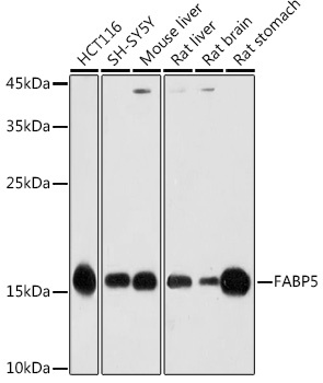
Western blot analysis of various lysates using FABP5 Rabbit pAb (CAB6373) at 1:1000 dilution. Secondary antibody: HRP-conjugated Goat anti-Rabbit IgG (H+L) (CABS014) at 1:10000 dilution. Lysates/proteins: 25μg per lane. Blocking buffer: 3% nonfat dry milk in TBST. Detection: ECL Basic Kit (AbGn00020). Exposure time: 30s.
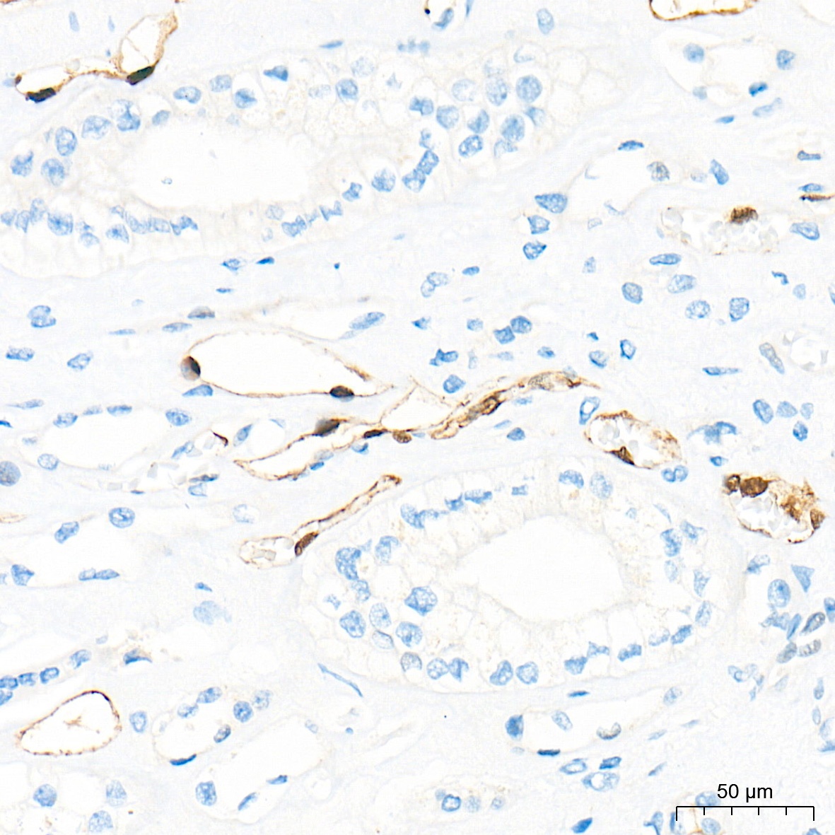
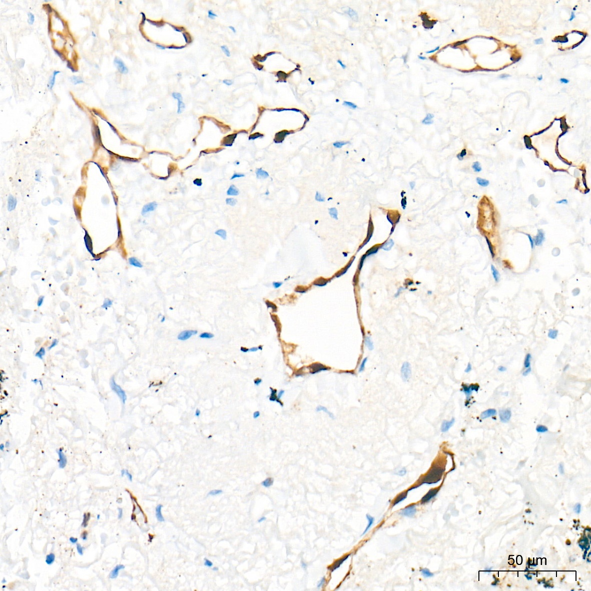
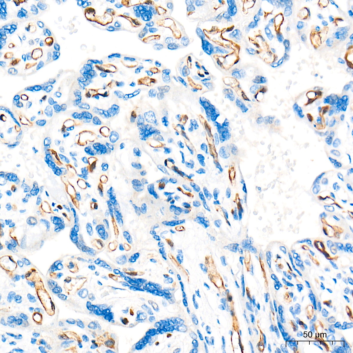
Immunohistochemistry analysis of paraffin-embedded Human kidney tissue using FABP5 Rabbit pAb (CAB6373) at a dilution of 1:300 (40x lens). High pressure antigen retrieval performed with 0.01M Tris-EDTA Buffer (pH 9.0) prior to IHC staining.
Immunohistochemistry analysis of paraffin-embedded Human lung tissue using FABP5 Rabbit pAb (CAB6373) at a dilution of 1:300 (40x lens). High pressure antigen retrieval performed with 0.01M Tris-EDTA Buffer (pH 9.0) prior to IHC staining.
Immunohistochemistry analysis of paraffin-embedded Human placenta tissue using FABP5 Rabbit pAb (CAB6373) at a dilution of 1:300 (40x lens). High pressure antigen retrieval performed with 0.01M Tris-EDTA Buffer (pH 9.0) prior to IHC staining.
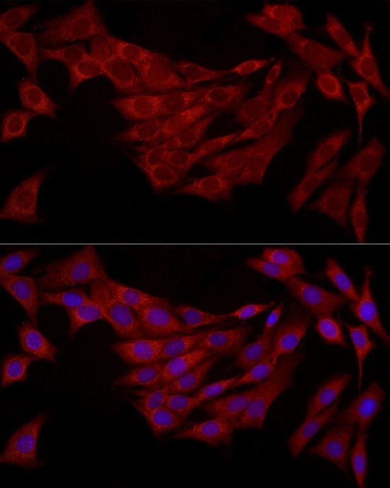
Immunofluorescence analysis of NIH/3T3 cells using FABP5 Rabbit pAb (CAB6373) at dilution of 1:200 (40x lens). Secondary antibody: Cy3-conjugated Goat anti-Rabbit IgG (H+L) (CABS007) at 1:500 dilution. Blue: DAPI for nuclear staining.
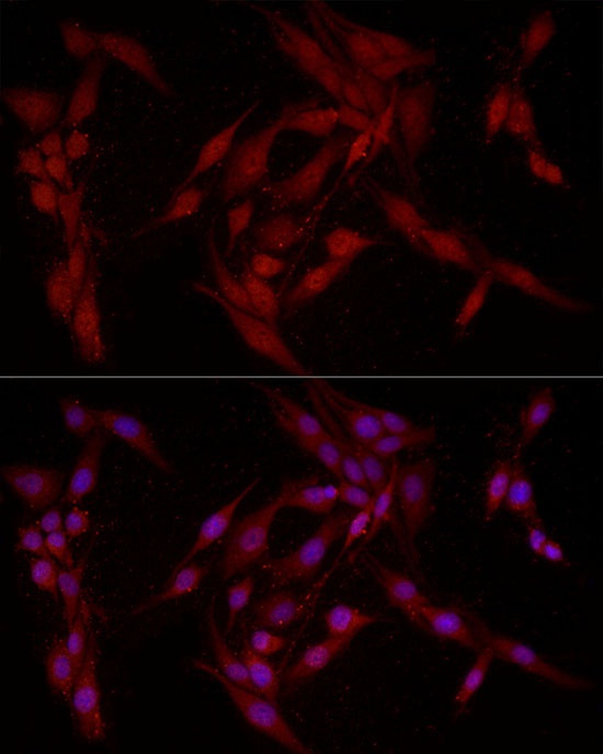
Immunofluorescence analysis of PC-12 cells using FABP5 Rabbit pAb (CAB6373) at dilution of 1:200 (40x lens). Secondary antibody: Cy3-conjugated Goat anti-Rabbit IgG (H+L) (CABS007) at 1:500 dilution. Blue: DAPI for nuclear staining.