The FAU Antibody (CAB12181) is a high-quality antibody developed for reliable detection and analysis of target proteins. This antibody, generated in rabbits, exhibits high reactivity with human samples and has been validated for use in Western blot applications. By binding specifically to the FAU protein, this antibody enables accurate detection and analysis in various cell types, making it an invaluable asset for studies in molecular biology and cancer research.The FAU protein, also known as Finkel-Biskis-Reilly murine sarcoma virus (FBR-MuSV) ubiquitously expressed oncogene homolog, plays a crucial role in regulating cell growth, proliferation, and apoptosis.
This antibody is validated for use in WB, ELISA applications and has demonstrated reactivity against Human, Mouse, Rat samples.
Product Name:
FAU Antibody
SKU:
CAB12181
Size:
20μL, 100μL
Reactivity:
Human, Mouse, Rat
Conjugate:
Unconjugated
Immunogen:
Recombinant protein (or fragment).This information is considered to be commercially sensitive.
This gene is the cellular homolog of the fox sequence in the Finkel-Biskis-Reilly murine sarcoma virus (FBR-MuSV). It encodes a fusion protein consisting of the ubiquitin-like protein fubi at the N terminus and ribosomal protein S30 at the C terminus. It has been proposed that the fusion protein is post-translationally processed to generate free fubi and free ribosomal protein S30. Fubi is a member of the ubiquitin family, and ribosomal protein S30 belongs to the S30E family of ribosomal proteins. Whereas the function of fubi is currently unknown, ribosomal protein S30 is a component of the 40S subunit of the cytoplasmic ribosome and displays antimicrobial activity. Pseudogenes derived from this gene are present in the genome. Similar to ribosomal protein S30, ribosomal proteins S27a and L40 are synthesized as fusion proteins with ubiquitin.
Purification Method
Affinity purification
Gene ID
2197
RRID
AB_2759068
Buffer Information
Store at -20℃. Avoid freeze / thaw cycles. Buffer: PBS with 0.01% thimerosal,50% glycerol,pH7.3.
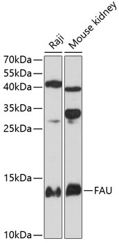
Western blot analysis of various lysates using FAU Rabbit pAb (CAB12181) at 1:3000 dilution. Secondary antibody: HRP-conjugated Goat anti-Rabbit IgG (H+L) (CABS014) at 1:10000 dilution. Lysates/proteins: 25μg per lane. Blocking buffer: 3% nonfat dry milk in TBST. Detection: ECL Basic Kit (AbGn00020). Exposure time: 90s.