The FGFR3 Monoclonal Antibody (CAB19052) is a high-quality antibody developed for reliable detection and analysis of target proteins. This antibody, produced in rabbits, exhibits high specificity and sensitivity for detecting FGFR3 in human samples, making it an ideal choice for Western blot applications.FGFR3 plays a crucial role in various biological processes, including skeletal development, bone formation, and cell proliferation. Dysregulation of FGFR3 has been linked to cancer and skeletal disorders, making it a significant target for therapeutic interventions.
This antibody is validated for use in IHC-P, ELISA applications and has demonstrated reactivity against Human samples.
Product Name:
FGFR3 Monoclonal Antibody
SKU:
CAB19052
Size:
20μL, 100μL
Reactivity:
Human
Clone Number:
ARC0398
Conjugate:
Unconjugated
Immunogen:
Synthetic peptide. This information is considered to be commercially sensitive.
Recommended starting concentration is 1 μg/mL. Please optimize the concentration based on your specific assay requirements.
Synonyms:
ACH, CEK2, JTK4, CD333, HSFGFR3EX, FGFR3
Cellular Localization:
Cell Membrane, Cytoplasmic Vesicle, Endoplasmic Reticulum, Secreted, Single-Pass Type I Membrane Protein.
Calculated MW:
88kDa
Observed MW:
RefertoFigures
This gene encodes a member of the fibroblast growth factor receptor (FGFR) family, with its amino acid sequence being highly conserved between members and among divergent species. FGFR family members differ from one another in their ligand affinities and tissue distribution. A full-length representative protein would consist of an extracellular region, composed of three immunoglobulin-like domains, a single hydrophobic membrane-spanning segment and a cytoplasmic tyrosine kinase domain. The extracellular portion of the protein interacts with fibroblast growth factors, setting in motion a cascade of downstream signals, ultimately influencing mitogenesis and differentiation. This particular family member binds acidic and basic fibroblast growth hormone and plays a role in bone development and maintenance. Mutations in this gene lead to craniosynostosis and multiple types of skeletal dysplasia.
Purification Method
Affinity purification
Gene ID
2261
RRID
AB_2862545
Buffer Information
Store at -20℃. Avoid freeze / thaw cycles. Buffer: PBS containing 50% glycerol and 0.05% BSA, preserved with proclin300 or sodium azide, pH 7.3.
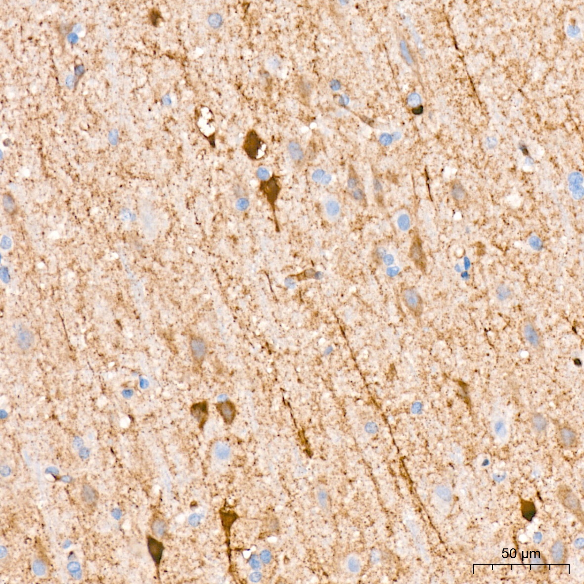
Immunohistochemistry analysis of paraffin-embedded Human brain tissue using FGFR3 Rabbit mAb (CAB19052) at a dilution of 1:2000 (40x lens). High pressure antigen retrieval was performed with 0.01 M citrate buffer (pH 6.0) prior to IHC staining.
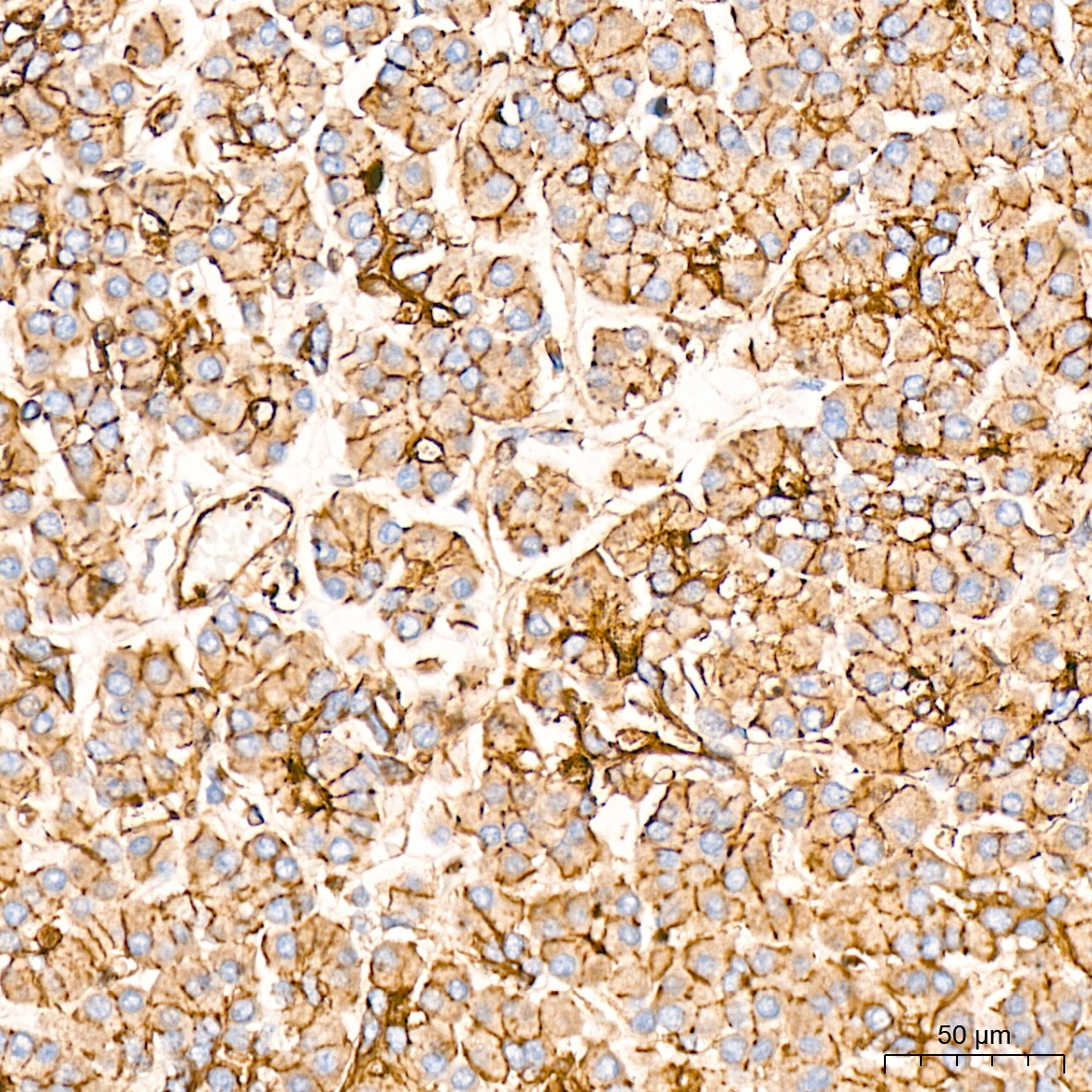
Immunohistochemistry analysis of paraffin-embedded Human pancreas tissue using FGFR3 Rabbit mAb (CAB19052) at a dilution of 1:2000 (40x lens). High pressure antigen retrieval was performed with 0.01 M citrate buffer (pH 6.0) prior to IHC staining.
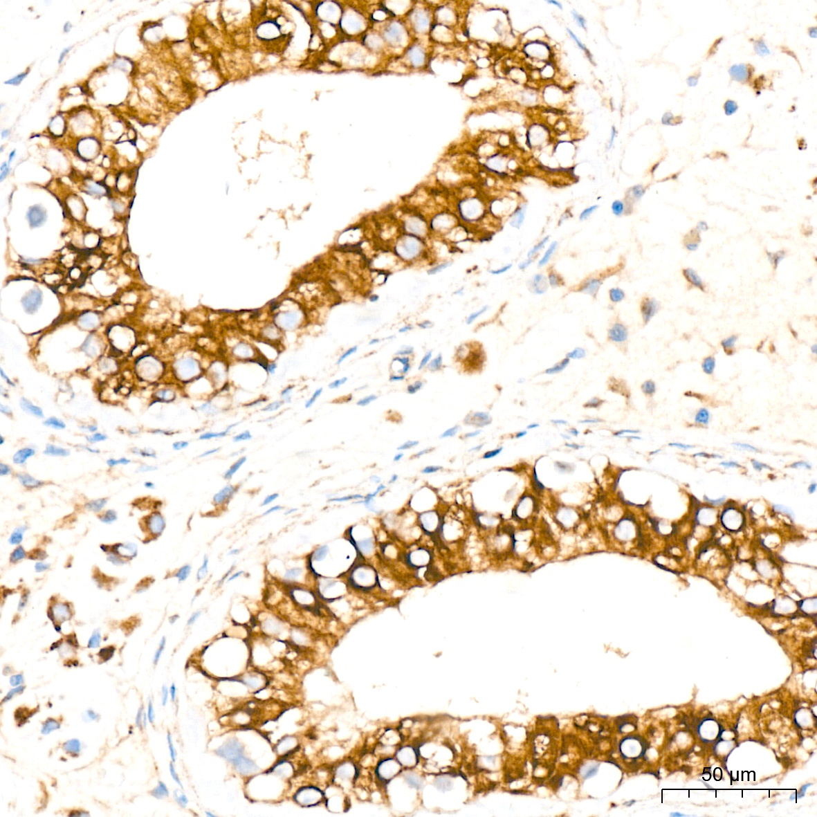
Immunohistochemistry analysis of paraffin-embedded Human testis tissue using FGFR3 Rabbit mAb (CAB19052) at a dilution of 1:2000 (40x lens). High pressure antigen retrieval was performed with 0.01 M citrate buffer (pH 6.0) prior to IHC staining.
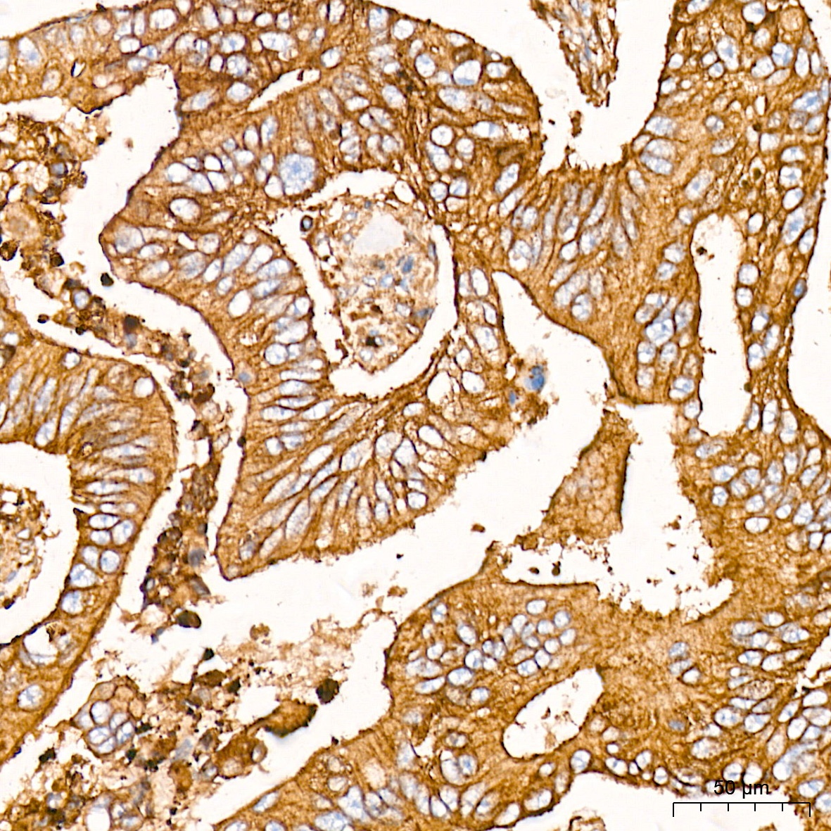
Immunohistochemistry analysis of paraffin-embedded Human colon carcinoma tissue using FGFR3 Rabbit mAb (CAB19052) at a dilution of 1:2000 (40x lens). High pressure antigen retrieval was performed with 0.01 M citrate buffer (pH 6.0) prior to IHC staining.


![Anti-FGFR3 [R09-1A7] Monoclonal Antibody (AGMB02866)](https://cdn11.bigcommerce.com/s-h68l9z2lnx/images/stencil/590x590/products/274155/679141/anti-fgfr3-r09-1a7-monoclonal-antibody-agmb02866__17656.1773037954.jpg?c=2 "Anti-FGFR3 [R09-1A7] Monoclonal Antibody (AGMB02866)")
![Anti-FGFR3 [R06-6F4] Monoclonal Antibody (AGMB02425)](https://cdn11.bigcommerce.com/s-h68l9z2lnx/images/stencil/590x590/products/273714/676341/anti-fgfr3-r06-6f4-monoclonal-antibody-agmb02425__64722.1773029171.jpg?c=2 "Anti-FGFR3 [R06-6F4] Monoclonal Antibody (AGMB02425)")

![Anti- FGFR3 [1B10] Monoclonal Antibody - Knockout Validated (AGMB06671)](https://cdn11.bigcommerce.com/s-h68l9z2lnx/images/stencil/590x590/products/277952/734185/anti-fgfr3-1b10-monoclonal-antibody-knockout-validated-agmb06671__81939.1777192396.jpg?c=2 "Anti- FGFR3 [1B10] Monoclonal Antibody - Knockout Validated (AGMB06671)")
![Anti-FGFR3 [R03-2C7] Monoclonal Antibody (AGMB02482)](https://cdn11.bigcommerce.com/s-h68l9z2lnx/images/stencil/590x590/products/273771/676474/anti-fgfr3-r03-2c7-monoclonal-antibody-agmb02482__95176.1773029546.jpg?c=2 "Anti-FGFR3 [R03-2C7] Monoclonal Antibody (AGMB02482)")