FSD1 Antibody is a premium polyclonal that offers outstanding performance and reliability for demanding research applications. Rigorously validated for ELISA, IHC, this antibody ensures consistent, reproducible results across multiple experimental platforms. Demonstrates excellent reactivity with Human samples, providing researchers with confidence in cross-species compatibility. Conveniently packaged in 50ug format to meet your experimental needs. For optimal performance, store at -20°C or -80°C and maintains stability for 12 months. Backed by rigorous quality control testing to ensure superior performance in your critical research applications.
Product Name:
FSD1 Antibody
SKU:
PACO60324
Size:
50μg
Isotype:
IgG
Host Species:
Rabbit
Reactivity:
Human
Immunogen:
Recombinant Human Fibronectin type III and SPRY domain-containing protein 1 protein (251-370AA)
Immunogen Species:
Homo sapiens (Human)
Uniprot No:
Q9BTV5
Form:
Liquid
Tested Applications:
ELISAIHC
Recommended Dilution:
Application
Recommended Dilution
IHC
1:500-1:1000
Synonyms:
Fibronectin type 3 and SPRY (spla, ryanodine) domain containing (with coiled-coil motif) 1 antibody, Fibronectin type 3 and SPRY domain containing 1 antibody, Fibronectin type III and SPRY domain containing 1 antibody, Fibronectin type III and SPRY domain-containing protein 1 antibody, Fibronectin type III and SPRY domains containing protein 1 antibody, FSD1 antibody, FSD1_HUMAN antibody, GLFND antibody, MGC3213 antibody, MGC58341 antibody, Microtubule-associated protein GLFND antibody, MID1-related protein 1 antibody, midline 1-related protein 1 antibody, MIR1 antibody, VLP27 antibody
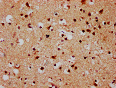
IHC image of PACO60324 diluted at 1:500 and staining in paraffin-embedded human brain tissue performed on a Leica BondTM system. After dewaxing and hydration, antigen retrieval was mediated by high pressure in a citrate buffer (pH 6.0). Section was blocked with 10% normal goat serum 30min at RT. Then primary antibody (1% BSA) was incubated at 4°C overnight. The primary is detected by a biotinylated secondary antibody and visualized using an HRP conjugated SP system.
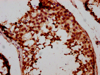
IHC image of PACO60324 diluted at 1:500 and staining in paraffin-embedded human testis tissue performed on a Leica BondTM system. After dewaxing and hydration, antigen retrieval was mediated by high pressure in a citrate buffer (pH 6.0). Section was blocked with 10% normal goat serum 30min at RT. Then primary antibody (1% BSA) was incubated at 4°C overnight. The primary is detected by a biotinylated secondary antibody and visualized using an HRP conjugated SP system.