The Gelsolin Antibody (CAB16869) is a high-quality antibody developed for reliable detection and analysis of target proteins. Raised in rabbits, this antibody is highly specific and reactive with human samples, making it ideal for use in Western blot applications. By binding to GSN, the antibody enables the detection and analysis of this important protein in various cell types.GSN, also known as gelsolin, is a key regulator of actin dynamics and is involved in a wide range of cellular processes, including cell motility, cytokinesis, and apoptosis. Dysregulation of GSN expression has been implicated in a variety of diseases, including cancer, neurodegenerative disorders, and autoimmune diseases.
This antibody is validated for use in WB, IHC-P, IF/ICC, ELISA applications and has demonstrated reactivity against Human, Mouse, Rat samples.
Product Name:
Gelsolin Antibody
SKU:
CAB16869
Size:
20μL, 100μL
Reactivity:
Human, Mouse, Rat
Immunogen:
Recombinant protein (or fragment).This information is considered to be commercially sensitive.
Recommended starting concentration is 1 μg/mL. Please optimize the concentration based on your specific assay requirements.
Synonyms:
ADF, AGEL, Gelsolin
Positive Sample:
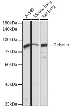
A-549, Mouse lung, Rat lung
Cellular Localization:
Cytoplasm, Secreted, Cytoskeleton.
Calculated MW:
86kDa
Observed MW:
86kDa
The protein encoded by this gene binds to the "plus" ends of actin monomers and filaments to prevent monomer exchange. The encoded calcium-regulated protein functions in both assembly and disassembly of actin filaments. Defects in this gene are a cause of familial amyloidosis Finnish type (FAF). Multiple transcript variants encoding several different isoforms have been found for this gene.
Purification Method
Affinity purification
Gene ID
2934
RRID
AB_2769700
Buffer Information
Store at -20℃. Avoid freeze / thaw cycles. Buffer: PBS containing 50% glycerol, preserved with proclin300 or sodium azide, pH 7.3.
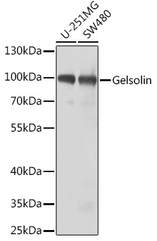
Western blot analysis of various lysates using Gelsolin Rabbit pAb (CAB16869) at 1:1000 dilution. Secondary antibody: HRP-conjugated Goat anti-Rabbit IgG (H+L) (CABS014) at 1:10000 dilution. Lysates/proteins: 25μg per lane. Blocking buffer: 3% nonfat dry milk in TBST. Detection: ECL Basic Kit (AbGn00020). Exposure time: 5s.
Western blot analysis of various lysates using Gelsolin Rabbit pAb (CAB16869) at 1:1000 dilution. Secondary antibody: HRP-conjugated Goat anti-Rabbit IgG (H+L) (CABS014) at 1:10000 dilution. Lysates/proteins: 25μg per lane. Blocking buffer: 3% nonfat dry milk in TBST. Detection: ECL Basic Kit (AbGn00020). Exposure time: 30s.
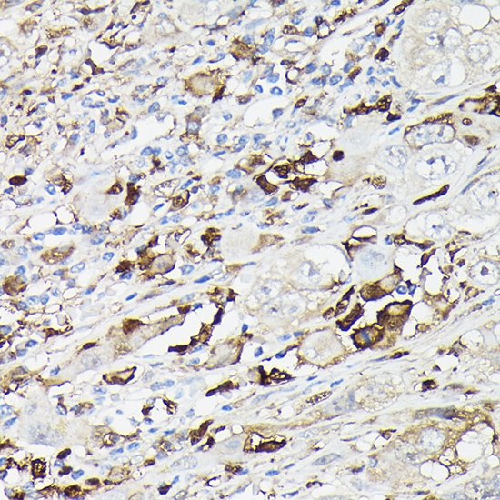
Immunohistochemistry analysis of paraffin-embedded Human liver cancer using Gelsolin Rabbit pAb (CAB16869) at dilution of 1:50 (40x lens). High pressure antigen retrieval performed with 0.01M Citrate buffer (pH 6.0) prior to IHC staining.
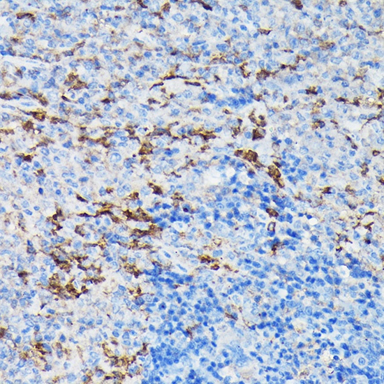
Immunohistochemistry analysis of paraffin-embedded Mouse spleen using Gelsolin Rabbit pAb (CAB16869) at dilution of 1:50 (40x lens). High pressure antigen retrieval performed with 0.01M Citrate buffer (pH 6.0) prior to IHC staining.
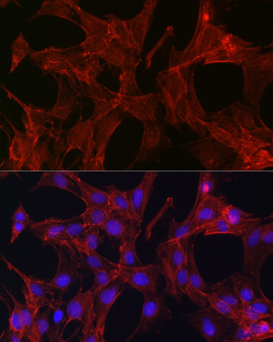
Immunofluorescence analysis of C6 cells using Gelsolin Rabbit pAb (CAB16869) at dilution of 1:50 (40x lens). Secondary antibody: Cy3-conjugated Goat anti-Rabbit IgG (H+L) (CABS007) at 1:500 dilution. Blue: DAPI for nuclear staining.
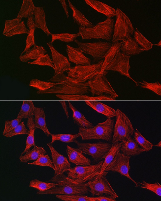
Immunofluorescence analysis of NIH-3T3 cells using Gelsolin Rabbit pAb (CAB16869) at dilution of 1:50 (40x lens). Secondary antibody: Cy3-conjugated Goat anti-Rabbit IgG (H+L) (CABS007) at 1:500 dilution. Blue: DAPI for nuclear staining.