The HDAC9 Monoclonal Antibody (CAB2226) is a high-quality antibody developed for reliable detection and analysis of target proteins. This antibody, generated in rabbits, exhibits high specificity and sensitivity for HDAC9 in human samples, making it ideal for use in Western blot and immunohistochemistry applications.HDAC9 plays a crucial role in the regulation of gene expression through the deacetylation of histone proteins, leading to changes in chromatin structure and transcriptional repression. Dysregulation of HDAC9 has been linked to various diseases, including cancer, cardiovascular disorders, and neurodegenerative diseases.
This antibody is validated for use in WB, IHC-P, IF/ICC, ELISA applications and has demonstrated reactivity against Human, Rat samples.
Product Name:
HDAC9 Monoclonal Antibody
SKU:
CAB2226
Size:
20μL, 100μL
Reactivity:
Human, Rat
Clone Number:
ARC0735
Conjugate:
Unconjugated
Immunogen:
Synthetic peptide. This information is considered to be commercially sensitive.
Histones play a critical role in transcriptional regulation, cell cycle progression, and developmental events. Histone acetylation/deacetylation alters chromosome structure and affects transcription factor access to DNA. The protein encoded by this gene has sequence homology to members of the histone deacetylase family. This gene is orthologous to the Xenopus and mouse MITR genes. The MITR protein lacks the histone deacetylase catalytic domain. It represses MEF2 activity through recruitment of multicomponent corepressor complexes that include CtBP and HDACs. This encoded protein may play a role in hematopoiesis. Multiple alternatively spliced transcripts have been described for this gene but the full-length nature of some of them has not been determined.
Purification Method
Affinity purification
Gene ID
9734
RRID
AB_2862978
Buffer Information
Store at -20℃. Avoid freeze / thaw cycles. Buffer: PBS containing 50% glycerol and 0.05% BSA, preserved with proclin300 or sodium azide, pH 7.3.
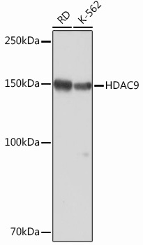
Western blot analysis of various lysates, using HDAC9 Rabbit mAb (CAB2226) at 1:1000 dilution. Secondary antibody: HRP-conjugated Goat anti-Rabbit IgG (H+L) (CABS014) at 1:10000 dilution. Lysates/proteins: 25μg per lane. Blocking buffer: 3% nonfat dry milk in TBST. Detection: ECL Basic Kit (AbGn00020). Exposure time: 3min.
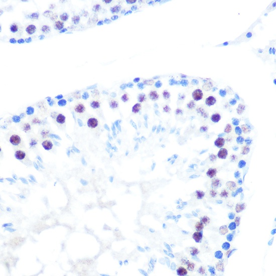
Immunohistochemistry analysis of paraffin-embedded Rat testis using HDAC9 Rabbit mAb (CAB2226) at dilution of 1:100 (40x lens). Microwave antigen retrieval performed with 0.01M PBS Buffer (pH 7.2) prior to IHC staining.
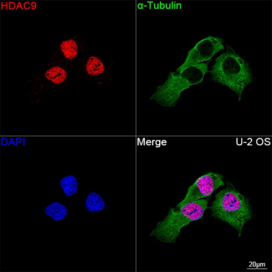
Confocal imaging of U-2 OS cells using HDAC9 Rabbit mAb (CAB2226, dilution 1:100) followed by a further incubation with Cy3 Goat Anti-Rabbit IgG (H+L) (CABS007, dilution 1:500) (Red). The cells were counterstained with α-Tubulin Mouse mAb (AC012, dilution 1:400) followed by incubation with ABflo® 488-conjugated Goat Anti-Mouse IgG (H+L) Ab (CABS076, dilution 1:500) (Green). DAPI was used for nuclear staining (Blue). Objective: 100x.
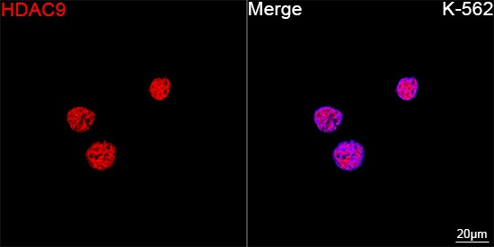
Confocal imaging of K-562 cells using HDAC9 Rabbit mAb (CAB2226, dilution 1:100) followed by a further incubation with Cy3 Goat Anti-Rabbit IgG (H+L) (CABS007,dilution 1:500) (Red). DAPI was used for nuclear staining (Blue). Objective: 100x.


![Anti-HDAC9 [R03-3C3] Monoclonal Antibody (AGMB00106)](https://cdn11.bigcommerce.com/s-h68l9z2lnx/images/stencil/590x590/products/271395/691515/anti-hdac9-r03-3c3-monoclonal-antibody-agmb00106__96042.1774503217.jpg?c=2 "Anti-HDAC9 [R03-3C3] Monoclonal Antibody (AGMB00106)")
![Anti-HDAC9 [R01-8I6] Monoclonal Antibody (AGMB00105)](https://cdn11.bigcommerce.com/s-h68l9z2lnx/images/stencil/590x590/products/271394/694197/anti-hdac9-r01-8i6-monoclonal-antibody-agmb00105__21560.1774511722.jpg?c=2 "Anti-HDAC9 [R01-8I6] Monoclonal Antibody (AGMB00105)")


