The HLA-DPB1 Antibody (CAB1412) is a high-quality antibody developed for reliable detection and analysis of target proteins. This antibody, generated in rabbits, is highly specific to human samples and is validated for use in various applications, including Western blot and immunohistochemistry.HLA-DPB1 plays a crucial role in presenting antigens to T cells and initiating immune responses. By targeting the HLA-DPB1 protein, researchers can gain insights into how the immune system recognizes and responds to foreign substances, offering important implications for studies in immunology, transplantation, infectious diseases, and autoimmunity.
This antibody is validated for use in WB, IHC-P, ELISA applications and has demonstrated reactivity against Human, Mouse, Rat samples.
Product Name:
HLA-DPB1 Antibody
SKU:
CAB1412
Size:
20μL, 100μL
Reactivity:
Human, Mouse, Rat
Conjugate:
Unconjugated
Immunogen:
Recombinant protein (or fragment).This information is considered to be commercially sensitive.
Recommended starting concentration is 1 μg/mL. Please optimize the concentration based on your specific assay requirements.
Synonyms:
DPB1, HLA-DP, HLA-DPB, HLA-DP1B, HLA-DPB1
Positive Sample:
Raji
Cellular Localization:
Cell Membrane, Endoplasmic Reticulum Membrane, Endosome Membrane, Golgi Apparatus, Lysosome Membrane, Single-Pass Type I Membrane Protein, Trans-Golgi Network Membrane.
Calculated MW:
29kDa
Observed MW:
25-35kDa
HLA-DPB belongs to the HLA class II beta chain paralogues. This class II molecule is a heterodimer consisting of an alpha (DPA) and a beta chain (DPB), both anchored in the membrane. It plays a central role in the immune system by presenting peptides derived from extracellular proteins. Class II molecules are expressed in antigen presenting cells (APC: B lymphocytes, dendritic cells, macrophages). The beta chain is approximately 26-28 kDa and its gene contains 6 exons. Exon one encodes the leader peptide, exons 2 and 3 encode the two extracellular domains, exon 4 encodes the transmembrane domain and exon 5 encodes the cytoplasmic tail. Within the DP molecule both the alpha chain and the beta chain contain the polymorphisms specifying the peptide binding specificities, resulting in up to 4 different molecules.
Purification Method
Affinity purification
Gene ID
3115
RRID
AB_2760976
Buffer Information
Store at -20℃. Avoid freeze / thaw cycles. Buffer: PBS containing 50% glycerol, preserved with proclin300 or sodium azide, pH 7.3.
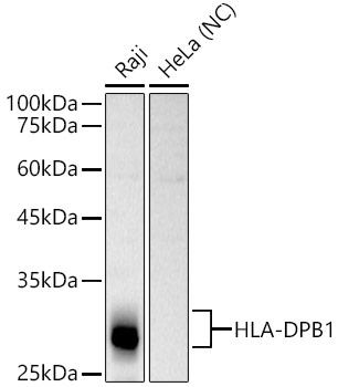
Western blot analysis of various lysates, using HLA-DPB1 Rabbit pAb (CAB1412) at 1:1000 dilution. Secondary antibody: HRP-conjugated Goat anti-Rabbit IgG (H+L) (CABS014) at 1:10000 dilution. Lysates/proteins: 25μg per lane. Blocking buffer: 3% nonfat dry milk in TBST. Detection: ECL Basic Kit (AbGn00020). Negative control (NC): HeLa Exposure time: 10s.
Immunohistochemistry analysis of paraffin-embedded Human lymphonodus using HLA-DPB1 Rabbit pAb (CAB1412) at dilution of 1:100 (40x lens). Microwave antigen retrieval performed with 0.01M PBS Buffer (pH 7.2) prior to IHC staining.
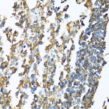
Immunohistochemistry analysis of paraffin-embedded Rat ovary using HLA-DPB1 Rabbit pAb (CAB1412) at dilution of 1:100 (40x lens). Microwave antigen retrieval performed with 0.01M PBS Buffer (pH 7.2) prior to IHC staining.
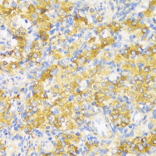
Immunohistochemistry analysis of paraffin-embedded Human lung cancer using HLA-DPB1 Rabbit pAb (CAB1412) at dilution of 1:100 (40x lens). Microwave antigen retrieval performed with 0.01M PBS Buffer (pH 7.2) prior to IHC staining.
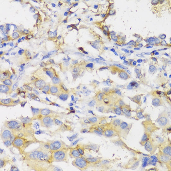
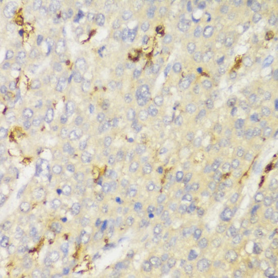
Immunohistochemistry analysis of paraffin-embedded Human liver cancer using HLA-DPB1 Rabbit pAb (CAB1412) at dilution of 1:100 (40x lens). Microwave antigen retrieval performed with 0.01M PBS Buffer (pH 7.2) prior to IHC staining.