The HRH3 Antibody (CAB16497) is a high-quality antibody developed for reliable detection and analysis of target proteins. This antibody is produced in rabbits and has been validated for use in Western blot applications, allowing for the detection and analysis of HRH3 protein in human samples. The histamine H3 receptor has been implicated in various physiological processes, including cognition, memory, and inflammation. Its dysregulation has been linked to neurological disorders, such as Alzheimer's disease and schizophrenia, making it a potential therapeutic target for drug development.
This antibody is validated for use in WB, IF/ICC, ELISA applications and has demonstrated reactivity against Human, Mouse, Rat samples.
Product Name:
HRH3 Antibody
SKU:
CAB16497
Size:
20μL, 100μL
Reactivity:
Human, Mouse, Rat
Conjugate:
Unconjugated
Immunogen:
Synthetic peptide. This information is considered to be commercially sensitive.
Recommended starting concentration is 1 μg/mL. Please optimize the concentration based on your specific assay requirements.
Synonyms:
HH3R, GPCR97, HRH3
Positive Sample:
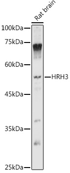
Rat brain
Cellular Localization:
Cell Membrane, Multi-Pass Membrane Protein.
Calculated MW:
49kDa
Observed MW:
49kDa
Histamine is a ubiquitous messenger molecule released from mast cells, enterochromaffin-like cells, and neurons. Its various actions are mediated by histamine receptors H1, H2, H3 and H4. This gene encodes one of the histamine receptors (H3) which belongs to the family 1 of G protein-coupled receptors. It is an integral membrane protein and can regulate neurotransmitter release. This receptor can also increase voltage-dependent calcium current in smooth muscles and innervates the blood vessels and the heart in cardiovascular system.
Purification Method
Affinity purification
Gene ID
11255
RRID
AB_2769844
Buffer Information
Store at -20℃. Avoid freeze / thaw cycles. Buffer: PBS containing 50% glycerol, preserved with proclin300 or sodium azide, pH 7.3.
Western blot analysis of lysates from Rat brain, using HRH3 Rabbit pAb (CAB16497) at 1:1000 dilution. Secondary antibody: HRP-conjugated Goat anti-Rabbit IgG (H+L) (CABS014) at 1:10000 dilution. Lysates/proteins: 25μg per lane. Blocking buffer: 3% nonfat dry milk in TBST. Detection: ECL Basic Kit (AbGn00020). Exposure time: 5s.
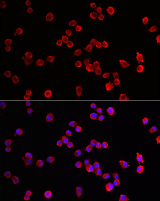
Immunofluorescence analysis of Neuro-2a cells using HRH3 Rabbit pAb (CAB16497) at dilution of 1:100 (40x lens). Secondary antibody: Cy3-conjugated Goat anti-Rabbit IgG (H+L) (CABS007) at 1:500 dilution. Blue: DAPI for nuclear staining.