The HUS1 Antibody (CAB13938) is a high-quality antibody developed for reliable detection and analysis of target proteins. This antibody, produced in rabbits, is highly specific to human samples and has been validated for use in Western blot applications. By binding to the Hus1 protein, this antibody allows for accurate detection and analysis in a variety of cell types, making it an essential tool for studies in genetics, molecular biology, and cancer research.Hus1 is a critical component of the Rad9-Hus1-Rad1 (9-1-1) complex, which plays a crucial role in DNA damage sensing and repair. Understanding the function and regulation of Hus1 is essential for unraveling the mechanisms underlying genomic stability and the development of cancer.
This antibody is validated for use in WB, IHC-P, ELISA applications and has demonstrated reactivity against Human, Mouse, Rat samples.
Product Name:
HUS1 Antibody
SKU:
CAB13938
Size:
20μL, 100μL
Reactivity:
Human, Mouse, Rat
Conjugate:
Unconjugated
Immunogen:
Recombinant protein (or fragment).This information is considered to be commercially sensitive.
Recommended starting concentration is 1 μg/mL. Please optimize the concentration based on your specific assay requirements.
Synonyms:
hHUS1, HUS1
Positive Sample:
Mouse kidney
Cellular Localization:
Cytoplasm, Nucleus.
Calculated MW:
32kDa
Observed MW:
32kDa
The protein encoded by this gene is a component of an evolutionarily conserved, genotoxin-activated checkpoint complex that is involved in the cell cycle arrest in response to DNA damage. This protein forms a heterotrimeric complex with checkpoint proteins RAD9 and RAD1. In response to DNA damage, the trimeric complex interacts with another protein complex consisting of checkpoint protein RAD17 and four small subunits of the replication factor C (RFC), which loads the combined complex onto the chromatin. The DNA damage induced chromatin binding has been shown to depend on the activation of the checkpoint kinase ATM, and is thought to be an early checkpoint signaling event. Alternative splicing results in multiple transcript variants.
Purification Method
Affinity purification
Gene ID
3364
RRID
AB_2760790
Buffer Information
Store at -20℃. Avoid freeze / thaw cycles. Buffer: PBS containing 50% glycerol, preserved with proclin300 or sodium azide, pH 7.3.
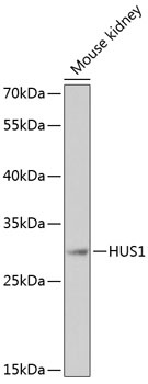
Western blot analysis of lysates from mouse kidney, using HUS1 Rabbit pAb (CAB13938) at 1:1000 dilution. Secondary antibody: HRP-conjugated Goat anti-Rabbit IgG (H+L) (CABS014) at 1:10000 dilution. Lysates/proteins: 25μg per lane. Blocking buffer: 3% nonfat dry milk in TBST. Detection: ECL Basic Kit (AbGn00020). Exposure time: 180s.
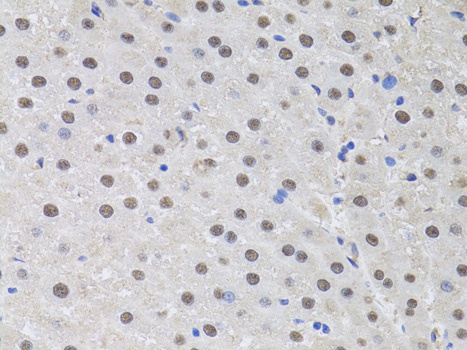
Immunohistochemistry analysis of paraffin-embedded Rat liver using HUS1 Rabbit pAb (CAB13938) at dilution of 1:100 (40x lens). Microwave antigen retrieval performed with 0.01M PBS Buffer (pH 7.2) prior to IHC staining.
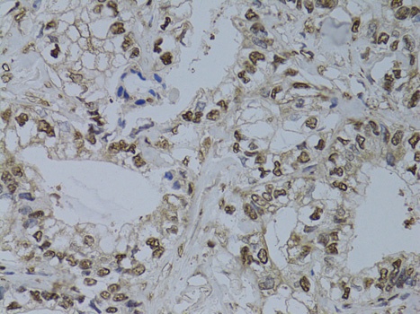
Immunohistochemistry analysis of paraffin-embedded Human gastric cancer using HUS1 Rabbit pAb (CAB13938) at dilution of 1:100 (40x lens). Microwave antigen retrieval performed with 0.01M PBS Buffer (pH 7.2) prior to IHC staining.
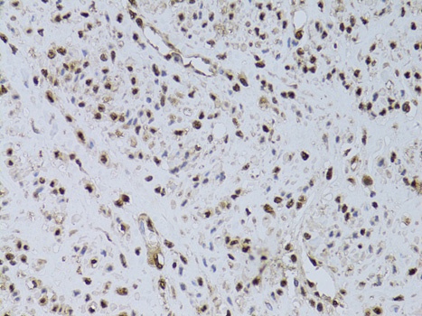
Immunohistochemistry analysis of paraffin-embedded Human uterine cancer using HUS1 Rabbit pAb (CAB13938) at dilution of 1:100 (40x lens). Microwave antigen retrieval performed with 0.01M PBS Buffer (pH 7.2) prior to IHC staining.