The HYAL1 Antibody (CAB6623) is a high-quality antibody developed for reliable detection and analysis of target proteins. This antibody, produced in rabbits, is highly specific to human samples and has been validated for use in Western blot applications. By binding to hyaluronidase-1, this antibody allows for the detection and analysis of this important enzyme in various cell types, making it an essential tool for studies in the fields of cell biology and cancer research.Hyaluronidase-1 plays a crucial role in tissue remodeling, inflammation, and cancer progression by breaking down hyaluronic acid, a major component of the extracellular matrix.
This antibody is validated for use in WB, IHC-P, IF/ICC, ELISA applications and has demonstrated reactivity against Human, Mouse, Rat samples.
Product Name:
HYAL1 Antibody
SKU:
CAB6623
Size:
20μL, 100μL
Reactivity:
Human, Mouse, Rat
Conjugate:
Unconjugated
Immunogen:
Recombinant protein (or fragment).This information is considered to be commercially sensitive.
Recommended starting concentration is 1 μg/mL. Please optimize the concentration based on your specific assay requirements.
Synonyms:
MPS9, NAT6, LUCA1, HYAL-1, HYAL1
Positive Sample:
PC-3
Cellular Localization:
Lysosome, Secreted.
Calculated MW:
48kDa
Observed MW:
48kDa
This gene encodes a lysosomal hyaluronidase. Hyaluronidases intracellularly degrade hyaluronan, one of the major glycosaminoglycans of the extracellular matrix. Hyaluronan is thought to be involved in cell proliferation, migration and differentiation. This enzyme is active at an acidic pH and is the major hyaluronidase in plasma. Mutations in this gene are associated with mucopolysaccharidosis type IX, or hyaluronidase deficiency. The gene is one of several related genes in a region of chromosome 3p21.3 associated with tumor suppression. Multiple transcript variants encoding different isoforms have been found for this gene.
Purification Method
Affinity purification
Gene ID
3373
RRID
AB_2767212
Buffer Information
Store at -20℃. Avoid freeze / thaw cycles. Buffer: PBS containing 50% glycerol, preserved with proclin300 or sodium azide, pH 7.3.
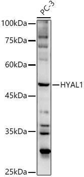
Western blot analysis of lysates from PC-3 cells, using HYAL1 Rabbit pAb (CAB6623) at 1:500 dilution. Secondary antibody: HRP-conjugated Goat anti-Rabbit IgG (H+L) (CABS014) at 1:10000 dilution. Lysates/proteins: 25μg per lane. Blocking buffer: 3% nonfat dry milk in TBST. Detection: ECL Basic Kit (AbGn00020). Exposure time: 180s.
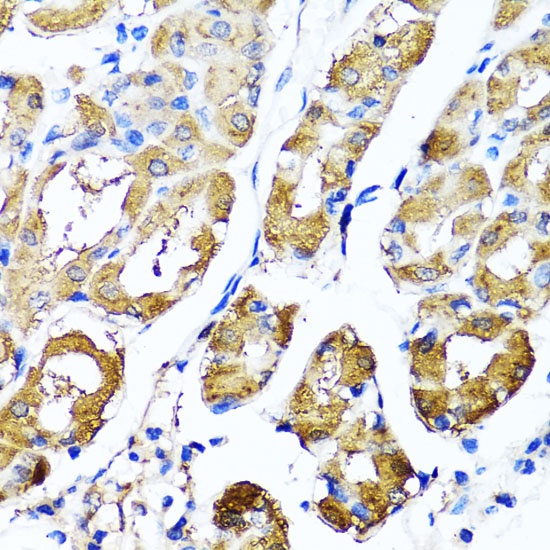
Immunohistochemistry analysis of paraffin-embedded Human stomach using HYAL1 Rabbit pAb (CAB6623) at dilution of 1:100 (40x lens). Microwave antigen retrieval performed with 0.01M PBS Buffer (pH 7.2) prior to IHC staining.
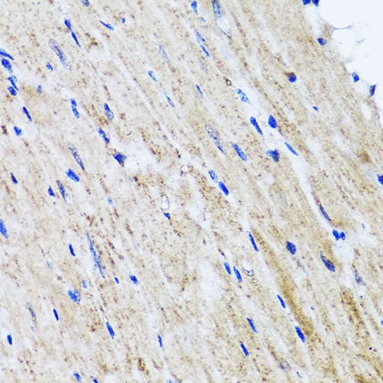
Immunohistochemistry analysis of paraffin-embedded Mouse heart using HYAL1 Rabbit pAb (CAB6623) at dilution of 1:100 (40x lens). Microwave antigen retrieval performed with 0.01M PBS Buffer (pH 7.2) prior to IHC staining.
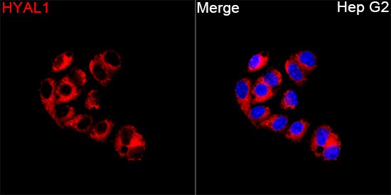
Immunofluorescence analysis of Hep G2 cells using HYAL1 Rabbit pAb (CAB6623) at a dilution of 1:100 (40x lens). Secondary antibody: Cy3-conjugated Goat anti-Rabbit IgG (H+L)(CABS007) at 1:500 dilution. Blue: DAPI for nuclear staining.