The CD11a/LFA-1A/ITGAL Antibody (CAB1644) is a high-quality antibody developed for reliable detection and analysis of target proteins. This antibody, generated in rabbits, specifically recognizes integrin alpha L in human samples and is validated for use in Western blot applications. By binding to integrin alpha L, this antibody allows for the detection and analysis of this important protein in various cell types, making it ideal for research in immunology and cancer.Integrin alpha L is a key player in immune cell trafficking and activation, playing a crucial role in inflammatory responses and immune cell function.
This antibody is validated for use in WB, IHC-P, IF/ICC, ELISA, IF-P applications and has demonstrated reactivity against Human, Mouse, Rat samples.
Product Name:
CD11a/LFA-1A/ITGAL Antibody
SKU:
CAB1644
Size:
20μL, 100μL
Reactivity:
Human, Mouse, Rat
Conjugate:
Unconjugated
Immunogen:
Recombinant protein (or fragment).This information is considered to be commercially sensitive.
Recommended starting concentration is 1 μg/mL. Please optimize the concentration based on your specific assay requirements.
Synonyms:
CD11A, LFA-1, LFA1A, CD11a/LFA-1A/ITGAL
Positive Sample:
Molt-4, Mouse spleen, Rat spleen
Cellular Localization:
Membrane, Single-Pass Type I Membrane Protein.
Calculated MW:
129kDa
Observed MW:
180kDa
ITGAL encodes the integrin alpha L chain. Integrins are heterodimeric integral membrane proteins composed of an alpha chain and a beta chain. This I-domain containing alpha integrin combines with the beta 2 chain (ITGB2) to form the integrin lymphocyte function-associated antigen-1 (LFA-1), which is expressed on all leukocytes. LFA-1 plays a central role in leukocyte intercellular adhesion through interactions with its ligands, ICAMs 1-3 (intercellular adhesion molecules 1 through 3), and also functions in lymphocyte costimulatory signaling. Two transcript variants encoding different isoforms have been found for this gene.
Purification Method
Affinity purification
Gene ID
3683
RRID
AB_2763701
Buffer Information
Store at -20℃. Avoid freeze / thaw cycles. Buffer: PBS containing 50% glycerol, preserved with proclin300 or sodium azide, pH 7.3.
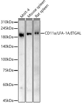
Western blot analysis of various lysates, using CD11a/LFA-1A/ITGAL Rabbit pAb (CAB1644) at 1:500 dilution. Secondary antibody: HRP-conjugated Goat anti-Rabbit IgG (H+L) (CABS014) at 1:10000 dilution. Lysates/proteins: 25μg per lane. Blocking buffer: 3% nonfat dry milk in TBST. Detection: ECL Basic Kit (AbGn00020). Exposure time: 180s.
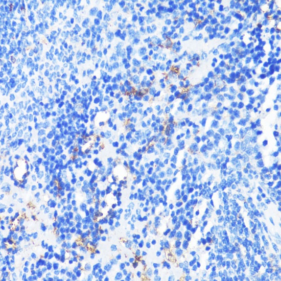
Immunohistochemistry analysis of paraffin-embedded Rat spleen using CD11a/LFA-1A/ITGAL Rabbit pAb (CAB1644) at dilution of 1:100 (40x lens). Microwave antigen retrieval performed with 0.01M PBS Buffer (pH 7.2) prior to IHC staining.
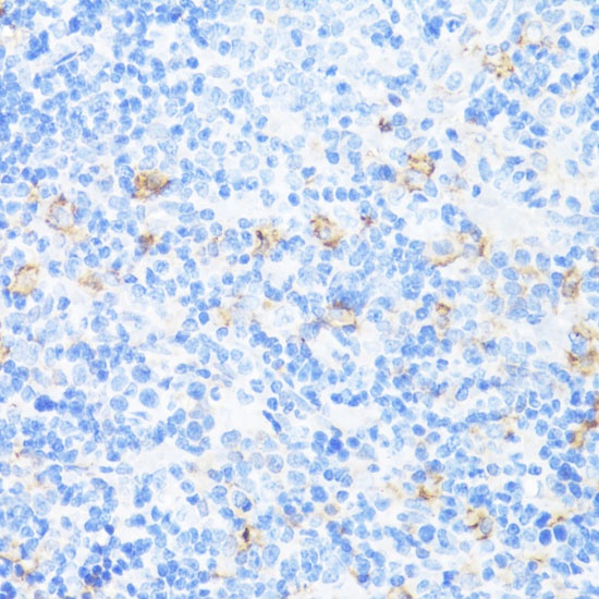
Immunohistochemistry analysis of paraffin-embedded Mouse spleen using CD11a/LFA-1A/ITGAL Rabbit pAb (CAB1644) at dilution of 1:100 (40x lens). Microwave antigen retrieval performed with 0.01M PBS Buffer (pH 7.2) prior to IHC staining.
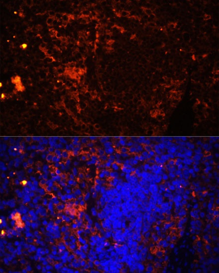
Immunofluorescence analysis of paraffin-embedded rat spleen using CD11a/LFA-1A/ITGAL Rabbit pAb (CAB1644) at dilution of 1:100 (40x lens). Secondary antibody: Cy3-conjugated Goat anti-Rabbit IgG (H+L) (CABS007) at 1:500 dilution. Blue: DAPI for nuclear staining.
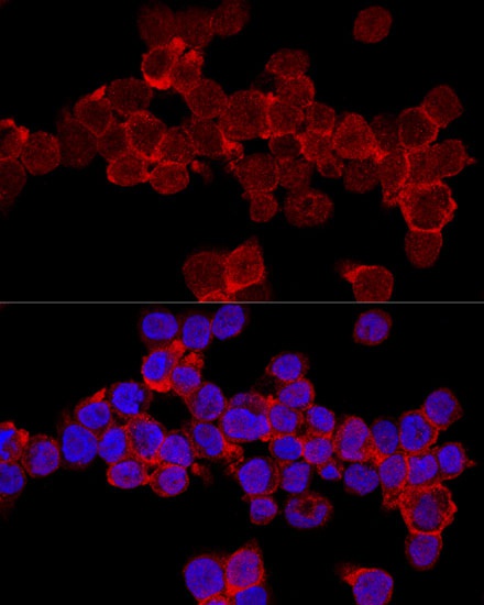
Confocal immunofluorescence analysis of THP-1 cells using CD11a/LFA-1A/ITGAL Rabbit pAb (CAB1644) at dilution of 1:200. Blue: DAPI for nuclear staining.


")

