The KLF4 Monoclonal Antibody (CAB13673) is a high-quality antibody developed for reliable detection and analysis of target proteins. This antibody, generated from rabbits, exhibits high affinity and specificity for human samples and has been rigorously validated for use in Western blot and immunohistochemistry applications.KLF4, also known as Krüppel-like factor 4, plays a crucial role in regulating gene expression and cell fate determination, making it a valuable target for investigating developmental biology, cancer, and stem cell research.
This antibody is validated for use in WB, IHC-P, IF/ICC, IP, ELISA applications and has demonstrated reactivity against Human, Mouse, Rat samples.
Product Name:
KLF4 Monoclonal Antibody
SKU:
CAB13673
Size:
20μL, 100μL
Reactivity:
Human, Mouse, Rat
Clone Number:
ARC0721
Conjugate:
Unconjugated
Immunogen:
A synthetic peptide corresponding to a sequence within amino acids 300-400 of human KLF4 (O43474).
0.5μg-4μg antibody for 200μg-400μg extracts of whole cells
ELISA
Recommended starting concentration is 1 μg/mL. Please optimize the concentration based on your specific assay requirements.
Synonyms:
EZF, GKLF, KLF4
Positive Sample:
A549, A-431, HeLa, Mouse lung
Cellular Localization:
Nucleus.
Calculated MW:
55kDa
Observed MW:
55kDa
This gene encodes a protein that belongs to the Kruppel family of transcription factors. The encoded zinc finger protein is required for normal development of the barrier function of skin. The encoded protein is thought to control the G1-to-S transition of the cell cycle following DNA damage by mediating the tumor suppressor gene p53. Mice lacking this gene have a normal appearance but lose weight rapidly, and die shortly after birth due to fluid evaporation resulting from compromised epidermal barrier function. Alternative splicing results in multiple transcript variants encoding different isoforms.
Purification Method
Affinity purification
Gene ID
9314
RRID
AB_2861693
Buffer Information
Store at -20℃. Avoid freeze / thaw cycles. Buffer: PBS containing 50% glycerol and 0.05% BSA, preserved with proclin300 or sodium azide, pH 7.3.
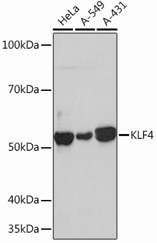
Western blot analysis of various lysates using KLF4 Rabbit mAb (CAB13673) at 1:1000 dilution. Secondary antibody: HRP-conjugated Goat anti-Rabbit IgG (H+L) (CABS014) at 1:10000 dilution. Lysates/proteins: 25μg per lane. Blocking buffer: 3% nonfat dry milk in TBST. Detection: ECL Basic Kit (AbGn00020). Exposure time: 30s.
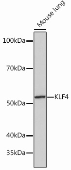
Western blot analysis of lysates from Mouse lung, using KLF4 Rabbit mAb (CAB13673) at 1:1000 dilution. Secondary antibody: HRP-conjugated Goat anti-Rabbit IgG (H+L) (CABS014) at 1:10000 dilution. Lysates/proteins: 25μg per lane. Blocking buffer: 3% nonfat dry milk in TBST. Detection: ECL Basic Kit (AbGn00020). Exposure time: 3min.
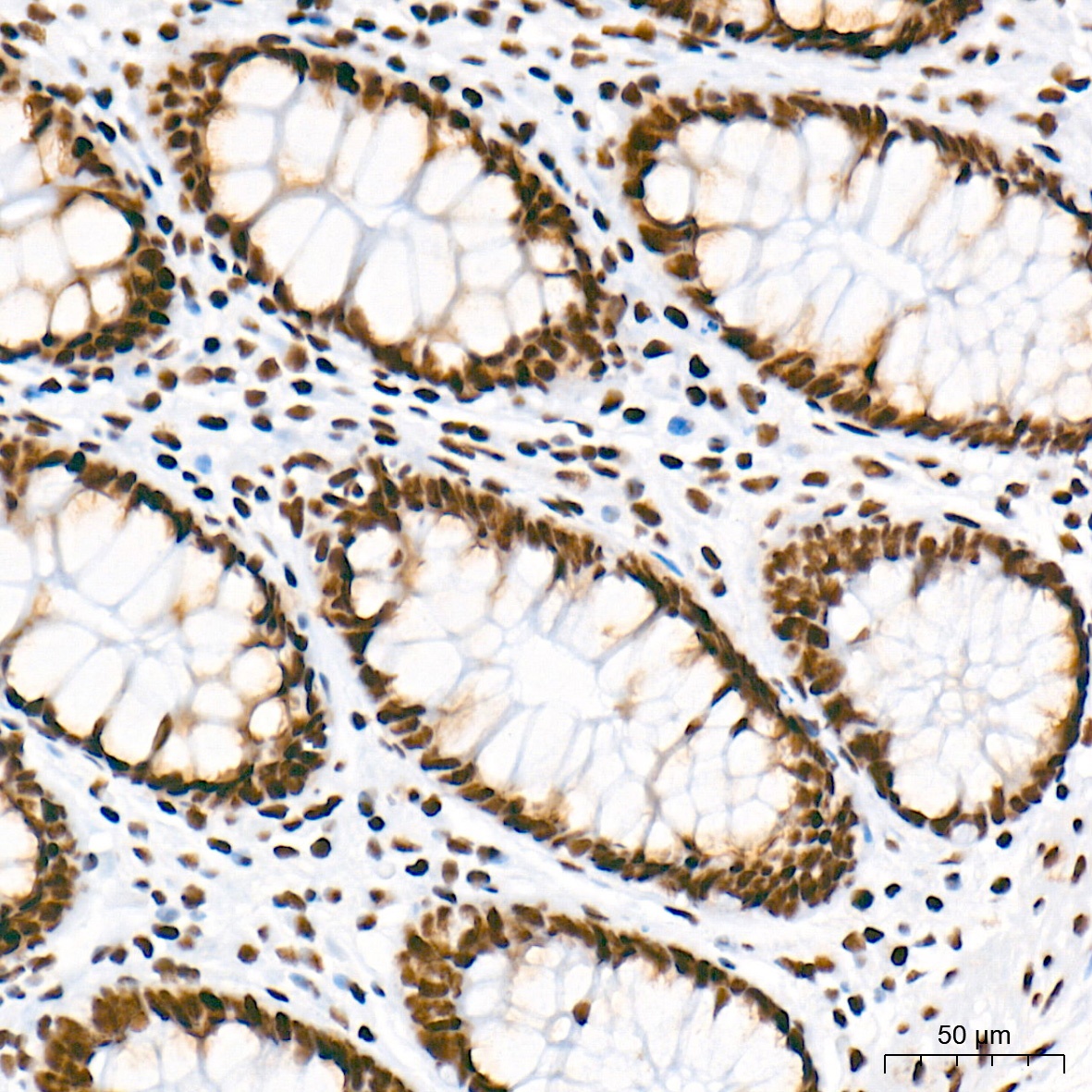
Immunohistochemistry analysis of paraffin-embedded Human colon tissue using KLF4 Rabbit mAb (CAB13673) at a dilution of 1:500 (40x lens). High pressure antigen retrieval performed with 0.01M Tris-EDTA Buffer (pH 9.0) prior to IHC staining.
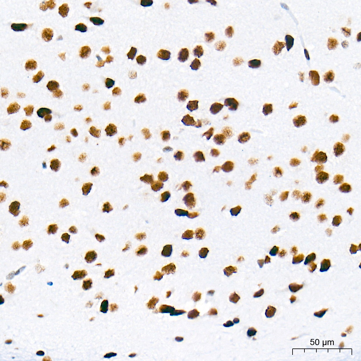
Immunohistochemistry analysis of paraffin-embedded Mouse brain tissue using KLF4 Rabbit mAb (CAB13673) at a dilution of 1:500 (40x lens). High pressure antigen retrieval performed with 0.01M Tris-EDTA Buffer (pH 9.0) prior to IHC staining.
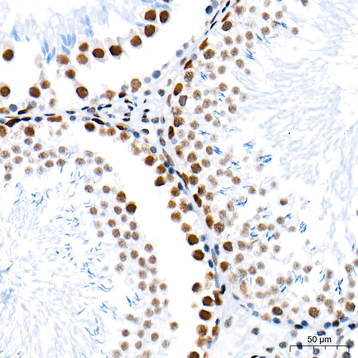
Immunohistochemistry analysis of paraffin-embedded Rat testis tissue using KLF4 Rabbit mAb (CAB13673) at a dilution of 1:500 (40x lens). High pressure antigen retrieval performed with 0.01M Tris-EDTA Buffer (pH 9.0) prior to IHC staining.
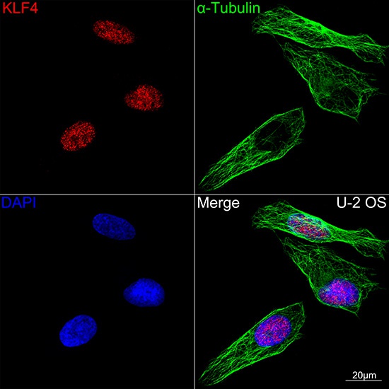
Confocal imaging of U-2 OS cells using KLF4 Rabbit mAb (CAB13673,dilution 1:100)(Red). The cells were counterstained with α-Tubulin Mouse mAb (AC012,dilution 1:400) (Green). DAPI was used for nuclear staining (blue). Objective: 100x.
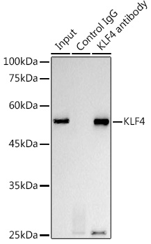
Immunoprecipitation analysis of 300 μg extracts from HeLa cells using 3 μg KLF4 Rabbit mAb (CAB13673). Western blot was performed from the immunoprecipitate using KLF4 Rabbit mAb (CAB13673) at a dilution of 1:1000.


![Anti-KLF4 [R03-5H7] Monoclonal Antibody (AGMB04150)](https://cdn11.bigcommerce.com/s-h68l9z2lnx/images/stencil/590x590/products/275439/679168/anti-klf4-r03-5h7-monoclonal-antibody-agmb04150__55793.1773038064.jpg?c=2 "Anti-KLF4 [R03-5H7] Monoclonal Antibody (AGMB04150)")
![Anti-KLF4 [R06-3M-1] Monoclonal Antibody (AGMB03653)](https://cdn11.bigcommerce.com/s-h68l9z2lnx/images/stencil/590x590/products/274942/680437/anti-klf4-r06-3m-1-monoclonal-antibody-agmb03653__42254.1773042037.jpg?c=2 "Anti-KLF4 [R06-3M-1] Monoclonal Antibody (AGMB03653)")
![Anti-KLF4 [R03-2I8] Monoclonal Antibody (AGMB00613)](https://cdn11.bigcommerce.com/s-h68l9z2lnx/images/stencil/590x590/products/271902/693029/anti-klf4-r03-2i8-monoclonal-antibody-agmb00613__26902.1774508008.jpg?c=2 "Anti-KLF4 [R03-2I8] Monoclonal Antibody (AGMB00613)")
![Anti-KLF4 [3B3-B11-E6] Monoclonal Antibody (AGMB04504)](https://cdn11.bigcommerce.com/s-h68l9z2lnx/images/stencil/590x590/products/275792/678940/anti-klf4-3b3-b11-e6-monoclonal-antibody-agmb04504__01043.1773037344.jpg?c=2 "Anti-KLF4 [3B3-B11-E6] Monoclonal Antibody (AGMB04504)")