The [KO Validated] MTH1 Antibody (CAB13330) is a high-quality antibody developed for reliable detection and analysis of target proteins. This antibody, generated in rabbits, is highly specific for human samples and has been rigorously validated for Western blot applications. By targeting the NUDT1 protein, this antibody enables precise detection and analysis in various cell types, making it ideal for investigations in DNA damage, cancer biology, and oxidative stress pathways.NUDT1, also known as nudix hydrolase 1, plays a crucial role in maintaining genomic integrity by hydrolyzing potentially mutagenic nucleotide metabolites. Its involvement in DNA repair processes and cellular defense against oxidative damage highlights its significance in cancer development and response to chemotherapy.
This antibody is validated for use in WB, IP, ELISA applications and has demonstrated reactivity against Human samples.
Product Name:
[KO Validated] MTH1 Antibody
SKU:
CAB13330
Size:
20μL, 100μL
Reactivity:
Human
Conjugate:
Unconjugated
Immunogen:
Recombinant protein (or fragment).This information is considered to be commercially sensitive.
0.5μg-4μg antibody for 200μg-400μg extracts of whole cells
ELISA
Recommended starting concentration is 1 μg/mL. Please optimize the concentration based on your specific assay requirements.
Synonyms:
MTH1, H1
Positive Sample:
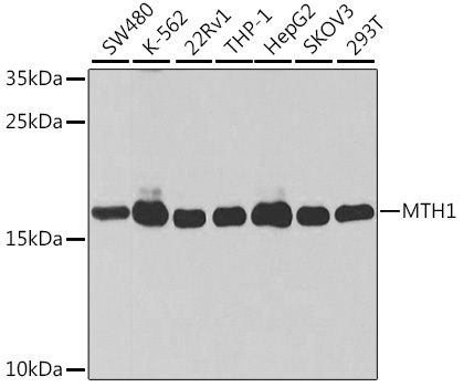
SW480, K562, 22RV1, THP-1, HepG2, SKOV3, 293T
Cellular Localization:
Cytoplasm, Mitochondrion Matrix, Nucleus.
Calculated MW:
18kDa
Observed MW:
18kDa
Misincorporation of oxidized nucleoside triphosphates into DNA/RNA during replication and transcription can cause mutations that may result in carcinogenesis or neurodegeneration. The protein encoded by this gene is an enzyme that hydrolyzes oxidized purine nucleoside triphosphates, such as 8-oxo-dGTP, 8-oxo-dATP, 2-hydroxy-dATP, and 2-hydroxy rATP, to monophosphates, thereby preventing misincorporation. The encoded protein is localized mainly in the cytoplasm, with some in the mitochondria, suggesting that it is involved in the sanitization of nucleotide pools both for nuclear and mitochondrial genomes. Several alternatively spliced transcript variants, some of which encode distinct isoforms, have been identified. Additional variants have been observed, but their full-length natures have not been determined. A rare single-nucleotide polymorphism that results in the production of an additional, longer isoform (p26) has been described.
Purification Method
Affinity purification
Gene ID
4521
RRID
AB_2861684
Buffer Information
Store at -20℃. Avoid freeze / thaw cycles. Buffer: PBS containing 50% glycerol, preserved with proclin300 or sodium azide, pH 7.3.
Western blot analysis of various lysates using [KO Validated] MTH1 Rabbit pAb (CAB13330) at 1:1000 dilution. Secondary antibody: HRP-conjugated Goat anti-Rabbit IgG (H+L) (CABS014) at 1:10000 dilution. Lysates/proteins: 25μg per lane. Blocking buffer: 3% nonfat dry milk in TBST. Detection: ECL Basic Kit (AbGn00020). Exposure time: 10s.
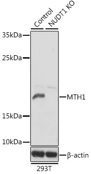
Western blot analysis of lysates from wild type (WT) and MTH1 knockout (KO) 293T cells, using [KO Validated] MTH1 Rabbit pAb (CAB13330) at 1:1000 dilution. Secondary antibody: HRP-conjugated Goat anti-Rabbit IgG (H+L) (CABS014) at 1:10000 dilution. Lysates/proteins: 25μg per lane. Blocking buffer: 3% nonfat dry milk in TBST. Detection: ECL Basic Kit (AbGn00020). Exposure time: 10s.