The MAP1LC3A Antibody (CAB11438) is a high-quality antibody developed for reliable detection and analysis of target proteins. This antibody, produced in rabbits, effectively recognizes the MAP1LC3A protein, a key player in autophagosome formation and maturation. Validated for use in Western blot applications, this antibody provides reliable and specific detection of MAP1LC3A in various experimental settings.MAP1LC3A, a mammalian homolog of yeast Atg8, is essential for autophagy, a cellular process crucial for maintaining cellular homeostasis and responding to stressors. Dysregulation of autophagy has been implicated in various diseases, including neurodegenerative disorders and cancer.
This antibody is validated for use in WB, IHC-P, ELISA applications and has demonstrated reactivity against Mouse, Rat samples.
Product Name:
MAP1LC3A Antibody
SKU:
CAB11438
Size:
20μL, 100μL
Reactivity:
Mouse, Rat
Conjugate:
Unconjugated
Immunogen:
Synthetic peptide. This information is considered to be commercially sensitive.
Sequence:
MPSD RPFK QRRS FADR CKEV QQIR DQHP SKIP VIIE RYKG EKQL PVLD KTKF LVPD HVNM SELV KIIR RRLQ LNPT QAFF LLVN QHSM VSVS TPIA DIYE
Tested Applications:
WBIHC-PELISA
Recommended Dilution:
WB
1:500 - 1:2000
IHC-P
1:50 - 1:200
ELISA
Recommended starting concentration is 1 μg/mL. Please optimize the concentration based on your specific assay requirements.
MAP1A and MAP1B are microtubule-associated proteins which mediate the physical interactions between microtubules and components of the cytoskeleton. MAP1A and MAP1B each consist of a heavy chain subunit and multiple light chain subunits. The protein encoded by this gene is one of the light chain subunits and can associate with either MAP1A or MAP1B. Two transcript variants encoding different isoforms have been found for this gene. The expression of variant 1 is suppressed in many tumor cell lines, suggesting that may be involved in carcinogenesis.
Purification Method
Affinity purification
Gene ID
84557
RRID
AB_2758560
Buffer Information
Store at -20℃. Avoid freeze / thaw cycles. Buffer: PBS containing 50% glycerol, preserved with proclin300 or sodium azide, pH 7.3.
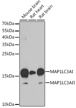
Western blot analysis of various lysates using MAP1LC3A Rabbit pAb (CAB11438) at 1:1000 dilution. Secondary antibody: HRP-conjugated Goat anti-Rabbit IgG (H+L) (CABS014) at 1:10000 dilution. Lysates/proteins: 25μg per lane. Blocking buffer: 3% nonfat dry milk in TBST. Detection: ECL Basic Kit (AbGn00020). Exposure time: 90s.
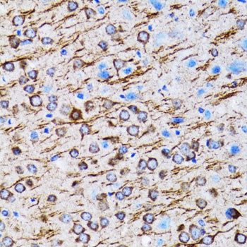
Immunohistochemistry analysis of paraffin-embedded Rat brain using MAP1LC3A Rabbit pAb (CAB11438) at dilution of 1:100 (40x lens). Microwave antigen retrieval performed with 0.01M PBS Buffer (pH 7.2) prior to IHC staining.