The MAP1LC3B Polyclonal Antibody (CAB21800) is a high-quality antibody developed for reliable detection and analysis of target proteins. This antibody, generated in rabbits, specifically targets the MAP1LC3B protein, a key player in autophagosome formation and autophagic flux.With its high reactivity to human samples, the MAP1LC3B Polyclonal Antibody is ideal for use in Western blotting applications, enabling researchers to easily detect and analyze MAP1LC3B levels in various cell types.
This antibody is validated for use in WB, ELISA applications and has demonstrated reactivity against Human, Rat samples.
Product Name:
MAP1LC3B Polyclonal Antibody
SKU:
CAB21800
Size:
20μL, 100μL
Reactivity:
Human, Rat
Conjugate:
Unconjugated
Immunogen:
Synthetic peptide. This information is considered to be commercially sensitive.
The product of this gene is a subunit of neuronal microtubule-associated MAP1A and MAP1B proteins, which are involved in microtubule assembly and important for neurogenesis. Studies on the rat homolog implicate a role for this gene in autophagy, a process that involves the bulk degradation of cytoplasmic component.
Purification Method
Affinity purification
Gene ID
81631
Buffer Information
Store at -20℃. Avoid freeze / thaw cycles. Buffer: PBS with 0.01% thimerosal,50% glycerol,pH7.3.
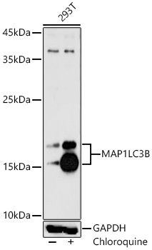
Western blot analysis of lysates from 293T cells using MAP1LC3B Rabbit pAb (CAB21800) at 1:1000 dilution. 293T cells were treated with Chloroquine (50 μM) at 37℃ for 20 hours. Secondary antibody: HRP-conjugated Goat anti-Rabbit IgG (H+L) (CABS014) at 1:10000 dilution. Lysates/proteins: 25 μg per lane. Blocking buffer: 3% nonfat dry milk in TBST. Detection: ECL Basic Kit (AbGn00020). Exposure time: 90s.
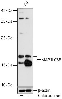
Western blot analysis of lysates from C6 cells using MAP1LC3B Rabbit pAb (CAB21800) at 1:1000 dilution. C6 cells were treated with Chloroquine (50 μM) at 37℃ for 20 hours. Secondary antibody: HRP-conjugated Goat anti-Rabbit IgG (H+L) (CABS014) at 1:10000 dilution. Lysates/proteins: 25 μg per lane. Blocking buffer: 3% nonfat dry milk in TBST. Detection: ECL Basic Kit (AbGn00020). Exposure time: 90s.
")
")