The [KO Validated] ERK2 Antibody (CAB11186) is a high-quality antibody developed for reliable detection and analysis of target proteins. This antibody, produced in rabbits, is highly specific to human samples and has been validated for use in Western blot and immunohistochemistry applications.MAPK1, also known as ERK2, is a member of the MAP kinase family that plays a critical role in various cellular processes, including proliferation, differentiation, and apoptosis. Dysregulation of the MAPK1 pathway has been implicated in a wide range of diseases, from cancer to neurodegenerative disorders.
This antibody is validated for use in WB, IHC-P, IF/ICC, ELISA applications and has demonstrated reactivity against Human, Mouse, Rat samples.
Product Name:
[KO Validated] ERK2 Antibody
SKU:
CAB11186
Size:
20μL, 100μL
Reactivity:
Human, Mouse, Rat
Conjugate:
Unconjugated
Immunogen:
Synthetic peptide. This information is considered to be commercially sensitive.
Sequence:
LNSK GYTK SIDI WSVG CILA EMLS NRPI FPGK HYLD QLNH ILGI LGSP SQED LNCI INLK ARNY LLSL PHKN KVPW NRLF PNAD SKAL DLLD KMLT FNPH K
Tested Applications:
WBIHC-PIF/ICCELISA
Recommended Dilution:
WB
1:500 - 1:1000
IHC-P
1:50 - 1:200
IF/ICC
1:50 - 1:200
ELISA
Recommended starting concentration is 1 μg/mL. Please optimize the concentration based on your specific assay requirements.
HeLa, A-549, NIH/3T3, C6, Mouse brain, Mouse thymus, Rat brain, HeLa, Mouse brain, Rat brain
Cellular Localization:
Cytoplasm, Nucleus, Centrosome, Cytoskeleton, Microtubule Organizing Center, Spindle.
Calculated MW:
41kDa
Observed MW:
42kDa
This gene encodes a member of the MAP kinase family. MAP kinases, also known as extracellular signal-regulated kinases (ERKs), act as an integration point for multiple biochemical signals, and are involved in a wide variety of cellular processes such as proliferation, differentiation, transcription regulation and development. The activation of this kinase requires its phosphorylation by upstream kinases. Upon activation, this kinase translocates to the nucleus of the stimulated cells, where it phosphorylates nuclear targets. One study also suggests that this protein acts as a transcriptional repressor independent of its kinase activity. The encoded protein has been identified as a moonlighting protein based on its ability to perform mechanistically distinct functions. Two alternatively spliced transcript variants encoding the same protein, but differing in the UTRs, have been reported for this gene.
Purification Method
Affinity purification
Gene ID
5594
RRID
AB_2814870
Buffer Information
Store at -20℃. Avoid freeze / thaw cycles. Buffer: PBS with 0.09% Sodium azide,50% glycerol,pH7.3.
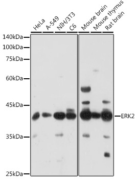
Western blot analysis of various lysates using [KO Validated] ERK2 Rabbit pAb (CAB11186) at 1:1000 dilution. Secondary antibody: HRP-conjugated Goat anti-Rabbit IgG (H+L) (CABS014) at 1:10000 dilution. Lysates/proteins: 25μg per lane. Blocking buffer: 3% nonfat dry milk in TBST. Detection: ECL Enhanced Kit (AbGn00021). Exposure time: 180s.
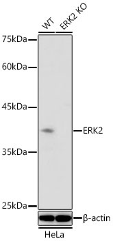
Western blot analysis of lysates from wild type (WT) and ERK2 knockout (KO) HeLa cells using [KO Validated] ERK2 Rabbit pAb (CAB11186) at 1:1000 dilution incubated overnight at 4℃. Secondary antibody: HRP-conjugated Goat anti-Rabbit IgG (H+L) (CABS014) at 1:10000 dilution. Lysates/proteins: 25 μg per lane. Blocking buffer: 3% nonfat dry milk in TBST. Detection: ECL Basic Kit (AbGn00020) Exposure time: 90 s.
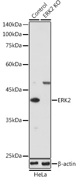
Western blot analysis of lysates from wild type (WT) and ERK2 knockout (KO) HeLa cells, using [KO Validated] ERK2 Rabbit pAb (CAB11186) at 1:1000 dilution. Secondary antibody: HRP-conjugated Goat anti-Rabbit IgG (H+L) (CABS014) at 1:10000 dilution. Lysates/proteins: 25μg per lane. Blocking buffer: 3% nonfat dry milk in TBST. Detection: ECL Enhanced Kit (AbGn00021). Exposure time: 180s.
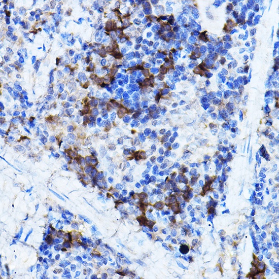
Immunohistochemistry analysis of paraffin-embedded Rat spleen using [KO Validated] ERK2 Rabbit pAb (CAB11186) at dilution of 1:100 (40x lens). High pressure antigen retrieval performed with 0.01M Citrate buffer (pH 6.0) prior to IHC staining.
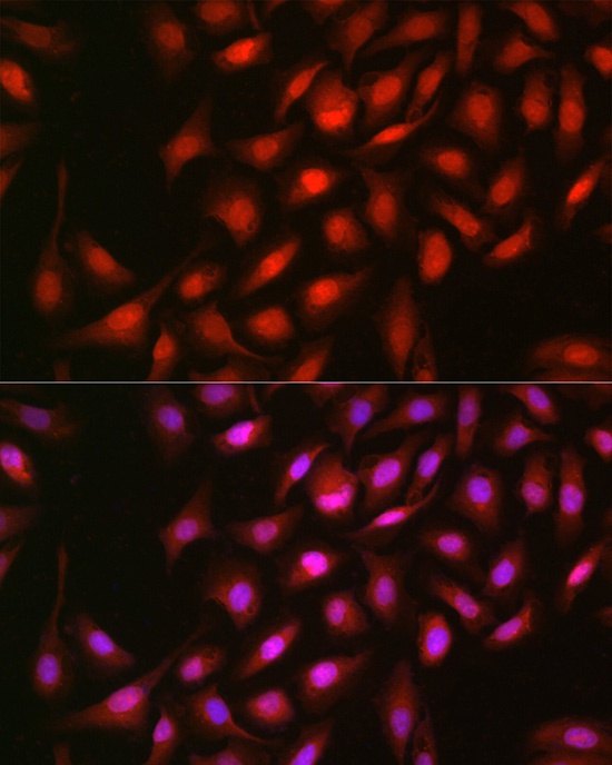
Immunofluorescence analysis of U-2 OS cells using [KO Validated] ERK2 Rabbit pAb (CAB11186) at dilution of 1:100 (40x lens). Secondary antibody: Cy3-conjugated Goat anti-Rabbit IgG (H+L) (CABS007) at 1:500 dilution. Blue: DAPI for nuclear staining.