The MATN4 Antibody (CAB2761) is a high-quality antibody developed for reliable detection and analysis of target proteins. This antibody, produced in rabbits, exhibits high reactivity with human samples and is validated for use in various applications including Western blot and immunohistochemistry.Matrilin-4 is known for its involvement in the formation and maintenance of cartilage tissues, making it a key target for studies in musculoskeletal biology and diseases such as osteoarthritis.
This antibody is validated for use in WB, ELISA applications and has demonstrated reactivity against Human, Mouse, Rat samples.
Product Name:
MATN4 Antibody
SKU:
CAB2761
Size:
20μL, 100μL
Reactivity:
Human, Mouse, Rat
Conjugate:
Unconjugated
Immunogen:
Recombinant protein (or fragment).This information is considered to be commercially sensitive.
Recommended starting concentration is 1 μg/mL. Please optimize the concentration based on your specific assay requirements.
Synonyms:
MATN4
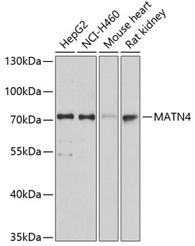
Positive Sample:
HepG2, NCI-H460, Mouse heart, Rat kidney
Cellular Localization:
Secreted.
Calculated MW:
68kDa
Observed MW:
73kDa
This gene encodes a member of von Willebrand factor A domain-containing protein family. The proteins of this family are thought to be involved in the formation of filamentous networks in the extracellular matrices of various tissues. This family member is thought to be play a role in reorganizing and regenerating the corneal matrix in granular and lattice type I dystrophies. It may also be involved in wound healing in the dentin-pulp complex. Alternative splicing results in multiple transcript variants.
Purification Method
Affinity purification
Gene ID
8785
RRID
AB_2764606
Buffer Information
Store at -20℃. Avoid freeze / thaw cycles. Buffer: PBS with 0.01% thimerosal,50% glycerol,pH7.3.
Western blot analysis of various lysates using MATN4 Rabbit pAb (CAB2761) at 1:3000 dilution. Secondary antibody: HRP-conjugated Goat anti-Rabbit IgG (H+L) (CABS014) at 1:10000 dilution. Lysates/proteins: 25μg per lane. Blocking buffer: 3% nonfat dry milk in TBST. Detection: ECL Basic Kit (AbGn00020). Exposure time: 10s.