The Monkey Calprotectin S100A8/S100A9 Heterodimer ELISA Kit is specifically designed for the accurate measurement of calprotectin S100A8/S100A9 levels in monkey serum, plasma, and tissue homogenate samples. This kit offers exceptional sensitivity and specificity, ensuring reliable and reproducible results for various research applications. Calprotectin S100A8/S100A9, also known as the heterodimer form of S100A8 and S100A9, is a key biomarker associated with inflammatory processes and immune response in various disease conditions.
Monitoring calprotectin levels can provide valuable insights into the pathogenesis of inflammatory disorders, autoimmune diseases, and cancer progression. With the Monkey Calprotectin S100A8/S100A9 Heterodimer ELISA Kit, researchers can accurately quantify calprotectin levels in monkey samples, facilitating the investigation of disease mechanisms and potential therapeutic targets. This kit is an essential tool for in-depth studies related to inflammation, immunity, and disease pathology in non-human primate models.
Serum, Plasma, Cell Culture Supernatant, Cell or Tissue Lysate, Other Liquid Samples
Storage:
2-8°C for 12 months.
Linearity:
Sample
1:2
1:4
1:8
Serum (n = 5)
92-104%
86-101%
81-98%
EDTA Plasma (n = 5)
87-101%
90-100%
83-98%
Heparin Plasma (n = 5)
89-103%
88-97%
80-93%
Recovery:
Sample
Recovery Range (%)
Average (%)
Serum (n = 5)
88-104
96
EDTA Plasma (n = 5)
87-104
95
Heparin Plasma (n = 5)
86-98
91
Note:The below protocol is a sample protocol. Protocols are specific to each batch/lot. For the correct instructions please follow the protocol included in your kit.
Step
Procedure
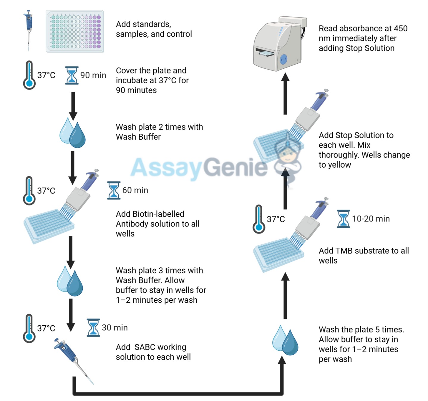
1
Reagent & Plate Preparation: Equilibrate reagents and TMB substrate to room temperature. Set standard, test sample and control (zero) wells on the pre-coated plate and record their positions.
2
Primary Incubation: Prepare standards, samples, blanks and load into designated wells. Incubate plate at 37°C for 90 minutes to allow antigen binding.
3
Detection Antibody Binding: Add biotin-labeled detection antibody and incubate at 37°C for 60 minutes.
4
HRP-Streptavidin Binding: Add HRP-Streptavidin (SABC) and incubate at 37°C for 30 minutes.
5
Color Development: Add TMB substrate and incubate in the dark for 10–20 minutes.
6
Stop Reaction & Reading: Add stop solution and measure absorbance at 450 nm immediately.
Sample Type
Protocol
Serum
Allow blood to clot, centrifuge at 1000 × g for 20 minutes, collect supernatant supernatant and store appropriately.
Plasma
Collect using anticoagulant tubes, centrifuge at 1000 × g for 15 minutes at 2–8°C and collect plasma.
Tissue Homogenate
Homogenize tissue in PBS with protease inhibitors, centrifuge and collect supernatant.
Cell Culture Supernatant
Centrifuge at 2500 rpm for 5 minutes and collect clarified supernatant.
Cell Lysate
Lyse cells using lysis buffer with protease inhibitors, centrifuge and collect protein supernatant.
Other Sample Types
For more information about how to process other sample types, (e.g., body fluids, breast milk & more), please contact our Tech Support Team at techsupport@assaygenie.com.
Component
Quantity
Storage
48T
96T
ELISA Microplate (Dismountable)
8×6
8×12
Place the test strips into a sealed foil bag with the desiccant. Store for 1 month at 2-8°C; Store for 12 months at -20°C.
Lyophilized Standard
1 vial
2 vial
Place the standards into a sealed foil bag with the desiccant. Store for 1 month at 2-8°C; Store for 12 months at -20°C.
Biotin-labeled Antibody (Lyophilized)
1 vial
1 vial
Place the standards into a sealed foil bag with the desiccant. Store for 1 month at 2-8°C; Store for 12 months at -20°C.
HRP-Streptavidin Conjugate (SABC, 100X)
60 ul
120 ul
2-8°C (Avoid direct light)
TMB Substrate
5 ml
10 ml
2-8°C (Avoid direct light)
Purified Water
200 ul
200 ul
2-8°C
Sample Dilution Buffer
10 ml
20 ml
2-8°C
Antibody Dilution Buffer
5 ml
10 ml
2-8°C
SABC Dilution Buffer
5 ml
10 ml
2-8°C
Stop Solution
5 ml
10 ml
2-8°C
Wash Buffer(25X)
15 ml
30 ml
2-8°C
Plate Sealer
3 pieces
5 pieces
-
Technical Manual
1 copy
1 copy
-
Lafoux et al.
Expansion of myeloid suppressor cells and suppression of Lassa virus-specific T cells during fatal Lassa fever
 ELISA Kit (AEFI00469)")
 ELISA Kit (AEFI00469)")
 ELISA Kit (AEFI00469)")
 ELISA Kit (AEFI00469)")
 ELISA Kit (AEFI03577)")
 ELISA Kit (AEFI03577)")
")


 ELISA Kit (MOFI01283)")