Mouse Complement C3 / C3 ELISA Kit
- SKU:
- MOFI00180
- Product Type:
- ELISA Kit
- Size:
- 96 Assays
- Uniprot:
- P01027
- Sensitivity:
- 1.875ng/ml
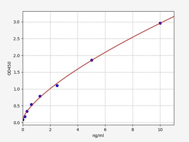
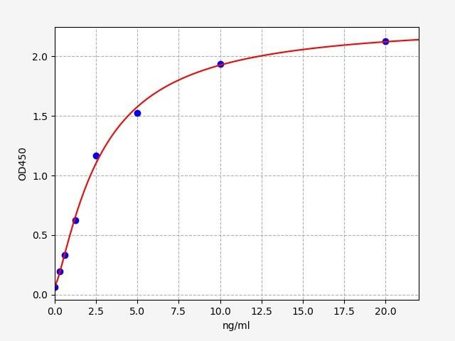
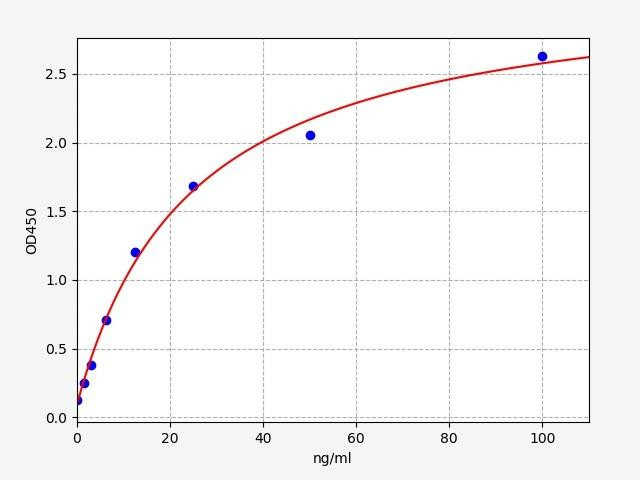
- Range:
- 3.125-200ng/ml
- ELISA Type:
- Sandwich
- Synonyms:
- C3, ASP, CPAMD1, AHUS5, ARMD9, C3a, C3b
- Reactivity:
- Mouse
- Research Area:
- Immunology

Description
Mouse Complement C3 / C3 ELISA Kit
Complement C3, a pivotal protein within the complement system, plays a multifaceted role in inflammation, immune defense, and clearance of immune complexes. The Mouse Complement C3 ELISA Kit not only enables quantitative assessment of Complement C3 levels but also provides valuable insights into its modulation, allowing researchers to delve deeper into immunological complexities.
Key Features
| Save Time | Pre-coated 96 well plate | |
| Quick Start | Kit includes all necessary reagents | |
| Publication Ready | Reproducible and reliable results |
Overview
|
Product Name: |
Mouse Complement C3 / C3 ELISA Kit |
|
Product Code: |
MOFI00180 |
|
Size: |
96 Assays |
|
Alias: |
C3, ASP, CPAMD1, AHUS5, ARMD9, C3a, C3b |
|
Detection Method: |
Sandwich ELISA |
|
Application: |
This immunoassay kit allows for the in vitro quantitative determination of Mouse C3 concentrations in serum plasma and other biological fluids. |
|
Sensitivity: |
1.875ng/ml |
|
Range: |
3.125-200ng/ml |
|
Storage: |
4°C for 6 months |
|
Note: |
For Research Use Only |
Additional Information
|
Recovery |
Matrices listed below were spiked with certain level of Mouse C3 and the recovery rates were calculated by comparing the measured value to the expected amount of Mouse C3 in samples.
|
||||||||||||||||||||
|
Linearity: |
The linearity of the kit was assayed by testing samples spiked with appropriate concentration of Mouse C3 and their serial dilutions. The results were demonstrated by the percentage of calculated concentration to the expected.
|
||||||||||||||||||||
|
Intra Assay |
CV < 8% |
||||||||||||||||||||
|
Inter Assay |
CV < 10% |
Kit Components
| Component | Quantity | Storage |
|
ELISA Microplate (Dismountable) |
8x12 strips |
4°C for 6 months |
|
Lyophilized Standard |
2 |
4°C/ -20°C |
|
Sample/Standard Dlution Buffer |
20ml |
4°C |
|
Biotin-labeled Antibody (Concentrated) |
120ul |
4°C (Protection from light) |
|
Antibody Dilution Buffer |
10ml |
4°C |
|
HRP-Streptavidin Conjugate (SABC) |
120ul |
4°C (Protect from light) |
|
SABC Dilution Buffer |
10ml |
4°C |
|
TMB Substrate |
10ml |
4°C (Protection from light) |
|
Stop Solution |
10ml |
4°C |
|
Wash Buffer (25X) |
30ml |
4°C |
|
Plate Sealer |
5 |
- |
Other materials required:
- Microplate reader with 450 nm wavelength filter
- Multichannel Pipette, Pipette, microcentrifuge tubes and disposable pipette tips
- Incubator
- Deionized or distilled water
- Absorbent paper
- Buffer resevoir
Protocol
*Note: Protocols are specific to each batch/lot. For the exact instructions please follow the protocol included in your kit.
Before adding to wells, equilibrate the SABC working solution and TMB substrate for at least 30 min at 37°C. When diluting samples and reagents, they must be mixed completely and evenly. It is recommended to plot a standard curve for each test.
| Step | Procedure |
|
1. |
Set standard, test sample and control (zero) wells on the pre-coated plate respectively, and then, record their positions. It is recommended to measure each standard and sample in duplicate. Wash plate 2 times before adding standard, sample and control (zero) wells! |
|
2. |
Aliquot 0.1ml standard solutions into the standard wells. |
|
3. |
Add 0.1 ml of Sample / Standard dilution buffer into the control (zero) well. |
|
4. |
Add 0.1 ml of properly diluted sample ( Human serum, plasma, tissue homogenates and other biological fluids.) into test sample wells. |
|
5. |
Seal the plate with a cover and incubate at 37 °C for 90 min. |
|
6. |
Remove the cover and discard the plate content, clap the plate on the absorbent filter papers or other absorbent material. Do NOT let the wells completely dry at any time. Wash plate X2. |
|
7. |
Add 0.1 ml of Biotin- detection antibody working solution into the above wells (standard, test sample & zero wells). Add the solution at the bottom of each well without touching the side wall. |
|
8. |
Seal the plate with a cover and incubate at 37°C for 60 min. |
|
9. |
Remove the cover, and wash plate 3 times with Wash buffer. Let wash buffer rest in wells for 1 min between each wash. |
|
10. |
Add 0.1 ml of SABC working solution into each well, cover the plate and incubate at 37°C for 30 min. |
|
11. |
Remove the cover and wash plate 5 times with Wash buffer, and each time let the wash buffer stay in the wells for 1-2 min. |
|
12. |
Add 90 µl of TMB substrate into each well, cover the plate and incubate at 37°C in dark within 10-20 min. (Note: This incubation time is for reference use only, the optimal time should be determined by end user.) And the shades of blue can be seen in the first 3-4 wells (with most concentrated standard solutions), the other wells show no obvious color. |
|
13. |
Add 50 µl of Stop solution into each well and mix thoroughly. The color changes into yellow immediately. |
|
14. |
Read the O.D. absorbance at 450 nm in a microplate reader immediately after adding the stop solution. |
Sample Type
When carrying out an ELISA assay it is important to prepare your samples in order to achieve the best possible results. Below we have a list of procedures for the preparation of samples for different sample types.
| Sample Type | Protocol |
|
Serum |
If using serum separator tubes, allow samples to clot for 30 minutes at room temperature. Centrifuge for 10 minutes at 1,000x g. Collect the serum fraction and assay promptly or aliquot and store the samples at -80°C. Avoid multiple freeze-thaw cycles. If serum separator tubes are not being used, allow samples to clot overnight at 2-8°C. Centrifuge for 10 minutes at 1,000x g. Remove serum and assay promptly or aliquot and store the samples at -80°C. Avoid multiple freeze-thaw cycles. |
|
Plasma |
Collect plasma using EDTA or heparin as an anticoagulant. Centrifuge samples at 4°C for 15 mins at 1000 × g within 30 mins of collection. Collect the plasma fraction and assay promptly or aliquot and store the samples at -80°C. Avoid multiple freeze-thaw cycles. Note: Over haemolysed samples are not suitable for use with this kit. |
|
Urine & Cerebrospinal Fluid |
Collect the urine (mid-stream) in a sterile container, centrifuge for 20 mins at 2000-3000 rpm. Remove supernatant and assay immediately. If any precipitation is detected, repeat the centrifugation step. A similar protocol can be used for cerebrospinal fluid. |
|
Cell culture supernatant |
Collect the cell culture media by pipette, followed by centrifugation at 4°C for 20 mins at 1500 rpm. Collect the clear supernatant and assay immediately. |
|
Cell lysates |
Solubilize cells in lysis buffer and allow to sit on ice for 30 minutes. Centrifuge tubes at 14,000 x g for 5 minutes to remove insoluble material. Aliquot the supernatant into a new tube and discard the remaining whole cell extract. Quantify total protein concentration using a total protein assay. Assay immediately or aliquot and store at ≤ -20 °C. |
|
Tissue homogenates |
The preparation of tissue homogenates will vary depending upon tissue type. Rinse tissue with 1X PBS to remove excess blood & homogenize in 20ml of 1X PBS (including protease inhibitors) and store overnight at ≤ -20°C. Two freeze-thaw cycles are required to break the cell membranes. To further disrupt the cell membranes you can sonicate the samples. Centrifuge homogenates for 5 mins at 5000xg. Remove the supernatant and assay immediately or aliquot and store at -20°C or -80°C. |
|
Tissue lysates |
Rinse tissue with PBS, cut into 1-2 mm pieces, and homogenize with a tissue homogenizer in PBS. Add an equal volume of RIPA buffer containing protease inhibitors and lyse tissues at room temperature for 30 minutes with gentle agitation. Centrifuge to remove debris. Quantify total protein concentration using a total protein assay. Assay immediately or aliquot and store at ≤ -20 °C |
|
Breast Milk |
Collect milk samples and centrifuge at 10,000 x g for 60 min at 4°C. Aliquot the supernatant and assay. For long term use, store samples at -80°C. Minimize freeze/thaw cycles. |
Complement 3 (C3) Background
What is C3 Complement?
Complement component 3 (C3) is a crucial protein that plays a central role in the complement system, which is a part of the body's innate immune response. The complement system consists of a group of proteins that work together to eliminate pathogens, trigger inflammation, and assist in the removal of damaged or dead cells. C3 is one of the key components of this system, and its activation initiates a cascade of events that lead to the immune response.
C3 Structure
C3 is a large glycoprotein that circulates in the blood plasma as an inactive form. It consists of two polypeptide chains, α and β, which are connected by a disulfide bond. The α chain is responsible for most of the protein's functions, while the β chain helps with stability. C3 is synthesized mainly in the liver and is produced at a constant rate, providing a continuous supply for immune responses.
C3 Function
The primary function of C3 is to serve as a critical component in the complement system activation. When an infection or tissue damage occurs, C3 is activated through different pathways, including the classical pathway, alternative pathway, and lectin pathway. Once activated, C3 undergoes proteolytic cleavage by enzymes known as convertases, leading to the formation of two fragments: C3a and C3b.
C3a is an anaphylatoxin, which means it triggers inflammation by attracting immune cells to the site of infection or tissue injury. It also enhances vascular permeability, allowing immune cells to reach the affected area more easily. Additionally, C3a participates in the regulation of the adaptive immune response by influencing the function of T and B cells.
C3b, on the other hand, plays a crucial role in the opsonization process. It binds to the surface of pathogens, such as bacteria or viruses, and facilitates their recognition and uptake by phagocytic cells like macrophages and neutrophils. Furthermore, C3b is involved in the formation of the C5 convertase, which leads to the subsequent activation of the terminal complement pathway and the assembly of the membrane attack complex (MAC). The MAC is responsible for direct lysis of certain pathogens by creating pores in their membranes.
C3 Complement Levels
Alterations in the C3 protein or its activation can have significant clinical implications. Deficiencies in C3 can lead to an increased susceptibility to infections, particularly with encapsulated bacteria. In some cases, deficiencies in other complement components can also indirectly affect C3 function.
Moreover, excessive activation of the complement system can result in autoimmune diseases, such as systemic lupus erythematosus (SLE) and glomerulonephritis. In these conditions, the immune system mistakenly attacks the body's own cells and tissues, leading to inflammation and organ damage. Elevated levels of C3 and its fragments can often be observed in the blood of individuals with active autoimmune diseases.
Mouse C3 ELISA Kit FAQs
Q: What is the purpose of Mouse C3 ELISA Kits?
Mouse C3 ELISA Kits are specifically designed laboratory kits used for the quantitative measurement of complement component 3 (C3) levels in mouse samples. These kits provide researchers and scientists with a reliable and efficient method to assess the concentration of C3 protein in various biological samples, such as serum, plasma, or tissue lysates. By measuring C3 levels, researchers can investigate the activity and function of the complement system, monitor complement-mediated immune responses, and study the role of C3 in various disease models.
Q: What type of samples can be used with Mouse C3 ELISA Kits?
Mouse C3 ELISA Kits can be used with a variety of biological samples, including mouse serum, plasma, cell culture supernatants, or tissue lysates. The kits provide guidelines and recommendations for sample preparation, dilution, and storage to ensure accurate and reliable results. It is important to follow the instructions provided with the specific kit to achieve optimal performance.
Q: Is the Mouse C3 ELISA Kit cross-species compatible?
The Mouse C3 ELISA Kit is specifically designed and validated for the measurement of complement component 3 (C3) levels in mouse samples. It is optimized for detecting and quantifying mouse C3 protein. However, the kit may not be suitable for use with samples from other species due to potential differences in C3 protein sequences or epitope recognition. For studies involving other species, it is recommended to use a species-specific or a cross-reactive ELISA kit validated for the particular species of interest. Additionally, consulting the product manual or contacting the manufacturer can provide more information regarding the cross-species compatibility of the Mouse C3 ELISA Kit.
Q: Where can I find additional technical support or assistance with the kit?
For any technical inquiries or assistance regarding the kit, you can reach out to our team. They will be available to answer your questions and provide the necessary guidance to ensure a successful experiment.
Related Products


Porcine Complement C3 / C3 ELISA Kit

