The MSH6 Polyclonal Antibody (CAB21445) is a high-quality antibody developed for reliable detection and analysis of target proteins. This antibody, generated in rabbits, is highly specific to human samples and has been rigorously validated for Western blot applications. By binding to the MSH6 protein, this antibody allows for accurate detection and analysis in a variety of cell types, making it an essential tool for studies in genetics, cancer research, and DNA repair mechanisms.
This antibody is validated for use in WB, IHC-P, IF/ICC, ELISA applications and has demonstrated reactivity against Human, Mouse, Rat, MK samples.
Product Name:
MSH6 Polyclonal Antibody
SKU:
CAB21445
Size:
20μL, 100μL
Reactivity:
Human, Mouse, Rat, MK
Conjugate:
Unconjugated
Immunogen:
Synthetic peptide. This information is considered to be commercially sensitive.
This gene encodes a member of the DNA mismatch repair MutS family. In E. coli, the MutS protein helps in the recognition of mismatched nucleotides prior to their repair. A highly conserved region of approximately 150 aa, called the Walker-A adenine nucleotide binding motif, exists in MutS homologs. The encoded protein heterodimerizes with MSH2 to form a mismatch recognition complex that functions as a bidirectional molecular switch that exchanges ADP and ATP as DNA mismatches are bound and dissociated. Mutations in this gene may be associated with hereditary nonpolyposis colon cancer, colorectal cancer, and endometrial cancer. Transcripts variants encoding different isoforms have been described.
Purification Method
Affinity purification
Gene ID
2956
Buffer Information
Store at -20℃. Avoid freeze / thaw cycles. Buffer: PBS containing 50% glycerol, preserved with proclin300 or sodium azide, pH 7.3.
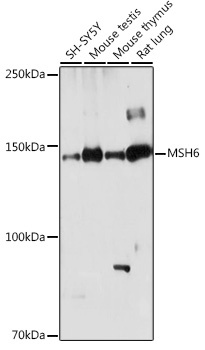
Western blot analysis of various lysates using MSH6 Rabbit pAb (CAB21445) at 1:1000 dilution. Secondary antibody: HRP-conjugated Goat anti-Rabbit IgG (H+L) (CABS014) at 1:10000 dilution. Lysates/proteins: 25μg per lane. Blocking buffer: 3% nonfat dry milk in TBST. Detection: ECL Basic Kit (AbGn00020). Exposure time: 180s.
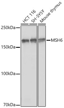
Western blot analysis of various lysates using [KO Validated] MSH6 Rabbit pAb (CAB21445) at 1:1000 dilution incubated overnight at 4℃. Secondary antibody: HRP-conjugated Goat anti-Rabbit IgG (H+L) (CABS014) at 1:10000 dilution. Lysates/proteins: 25 μg per lane. Blocking buffer: 3% nonfat dry milk in TBST. Detection: ECL Basic Kit (AbGn00020) Exposure time: 10 s.
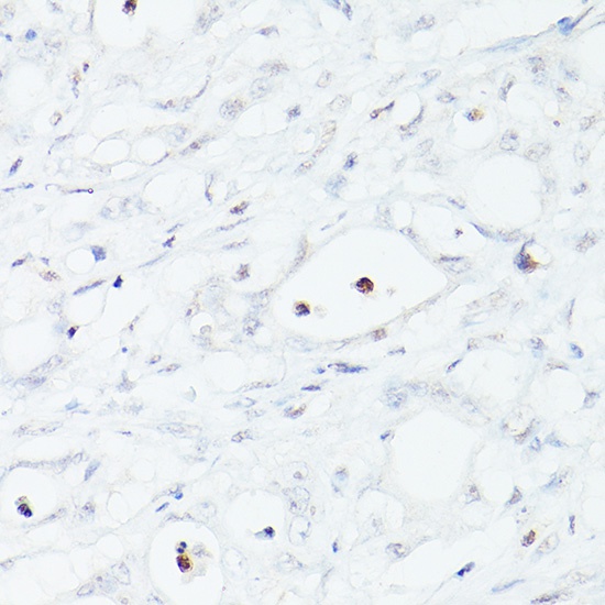
Immunohistochemistry analysis of paraffin-embedded Human colon cancer (loss of MSH6 expression) using MSH6 Rabbit pAb (CAB21445) at dilution of 1:200 (40x lens). High pressure antigen retrieval performed with 0.01M Citrate buffer (pH 6.0) prior to IHC staining.
Immunohistochemistry analysis of paraffin-embedded Human colon carcinoma using MSH6 Rabbit pAb (CAB21445) at dilution of 1:200 (40x lens). High pressure antigen retrieval performed with 0.01M Citrate buffer (pH 6.0) prior to IHC staining.
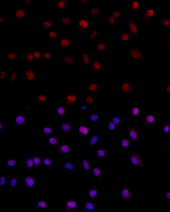
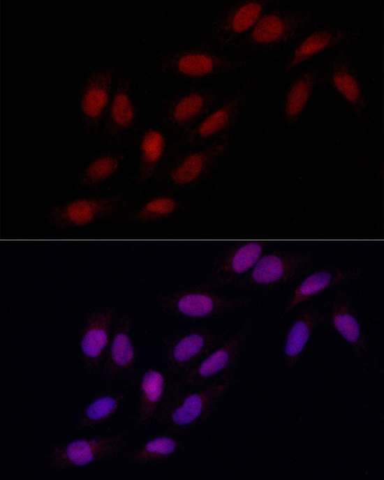
Immunofluorescence analysis of A-549 cells using MSH6 Rabbit pAb (CAB21445) at dilution of 1:200 (40x lens). Secondary antibody: Cy3-conjugated Goat anti-Rabbit IgG (H+L) (CABS007) at 1:500 dilution. Blue: DAPI for nuclear staining.
Immunofluorescence analysis of U2OS cells using MSH6 Rabbit pAb (CAB21445) at dilution of 1:200 (40x lens). Secondary antibody: Cy3-conjugated Goat anti-Rabbit IgG (H+L) (CABS007) at 1:500 dilution. Blue: DAPI for nuclear staining.
 at 1:1000 dilution. Secondary antibody: HRP Goat Anti-Rabbit IgG (H+L) at 1:10000 dilution. Lysates/proteins: 25μg per lane. Blocking buffer: 3% nonfat dry milk in TBST.")

 at 1:1000 dilution. Secondary antibody: HRP Goat Anti-Rabbit IgG (H+L) at 1:10000 dilution. Lysates/proteins: 25μg per lane. Blocking buffer: 3% nonfat dry milk in TBST.")

![Anti-MSH6 [R02-6G6] Monoclonal Antibody (AGMB01252)](https://cdn11.bigcommerce.com/s-h68l9z2lnx/images/stencil/590x590/products/272541/695257/anti-msh6-r02-6g6-monoclonal-antibody-agmb01252__90401.1774515077.jpg?c=2 "Anti-MSH6 [R02-6G6] Monoclonal Antibody (AGMB01252)")
![Anti-MSH6 [R05-2B5] Monoclonal Antibody (AGMB01251)](https://cdn11.bigcommerce.com/s-h68l9z2lnx/images/stencil/590x590/products/272540/694413/anti-msh6-r05-2b5-monoclonal-antibody-agmb01251__02877.1774512429.jpg?c=2 "Anti-MSH6 [R05-2B5] Monoclonal Antibody (AGMB01251)")
![Anti-MSH6 [R05-3F-5] Monoclonal Antibody (AGMB06333)](https://cdn11.bigcommerce.com/s-h68l9z2lnx/images/stencil/590x590/products/277614/732615/anti-msh6-r05-3f-5-monoclonal-antibody-agmb06333__55000.1777187378.jpg?c=2 "Anti-MSH6 [R05-3F-5] Monoclonal Antibody (AGMB06333)")
![Anti-MSH6 [7H4-4C1-9C5] Monoclonal Antibody (AGMB06132)](https://cdn11.bigcommerce.com/s-h68l9z2lnx/images/stencil/590x590/products/277413/678201/anti-msh6-7h4-4c1-9c5-monoclonal-antibody-agmb06132__42300.1773035052.jpg?c=2 "Anti-MSH6 [7H4-4C1-9C5] Monoclonal Antibody (AGMB06132)")