The NDUFB8 Monoclonal Antibody (CAB19732) is a high-quality antibody developed for reliable detection and analysis of target proteins. This monoclonal antibody, developed through immunization of mice, exhibits high specificity for human samples and has been validated for use in various applications, including Western blotting. By targeting the NDUFB8 protein, this antibody enables accurate detection and analysis of mitochondrial complex I activity in different cell types, making it an essential component for studies in bioenergetics, mitochondrial dysfunction, and related diseases.The NDUFB8 protein is essential for the proper function of complex I, which is a central player in cellular energy production through oxidative phosphorylation.
This antibody is validated for use in WB, IHC-P, IF/ICC, ELISA, IF-P applications and has demonstrated reactivity against Human, Mouse, Rat samples.
Product Name:
NDUFB8 Monoclonal Antibody
SKU:
CAB19732
Size:
20μL, 100μL
Reactivity:
Human, Mouse, Rat
Clone Number:
ARC2259
Conjugate:
Unconjugated
Immunogen:
Recombinant protein (or fragment).This information is considered to be commercially sensitive.
Involved in mitochondrial respiratory chain complex I assembly. Located in endoplasmic reticulum and mitochondrion. Part of mitochondrial respiratory chain complex I. Implicated in nuclear type mitochondrial complex I deficiency 32. Biomarker of Alzheimer's disease and Parkinson's disease.
Purification Method
Affinity purification
Gene ID
4714
RRID
AB_2938519
Buffer Information
Store at -20℃. Avoid freeze / thaw cycles. Buffer: PBS containing 50% glycerol and 0.05% BSA, preserved with proclin300 or sodium azide, pH 7.3.
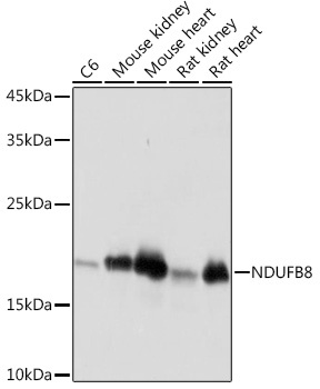
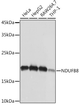
Western blot analysis of various lysates using NDUFB8 Rabbit mAb (CAB19732) at 1:1000 dilution. Secondary antibody: HRP-conjugated Goat anti-Rabbit IgG (H+L) (CABS014) at 1:10000 dilution. Lysates/proteins: 25μg per lane. Blocking buffer: 3% nonfat dry milk in TBST. Detection: ECL Basic Kit (AbGn00020). Exposure time: 1s.
Western blot analysis of various lysates using NDUFB8 Rabbit mAb (CAB19732) at 1:1000 dilution. Secondary antibody: HRP-conjugated Goat anti-Rabbit IgG (H+L) (CABS014) at 1:10000 dilution. Lysates/proteins: 25μg per lane. Blocking buffer: 3% nonfat dry milk in TBST. Detection: ECL Basic Kit (AbGn00020). Exposure time: 1s.
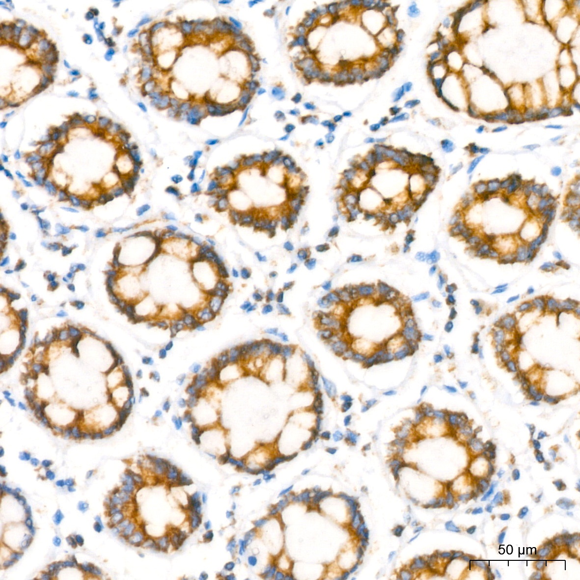
Immunohistochemistry analysis of paraffin-embedded Human colon using NDUFB8 Rabbit mAb (CAB19732) at dilution of 1:200 (40x lens). High pressure antigen retrieval performed with 0.01M Citrate buffer (pH 6.0) prior to IHC staining.
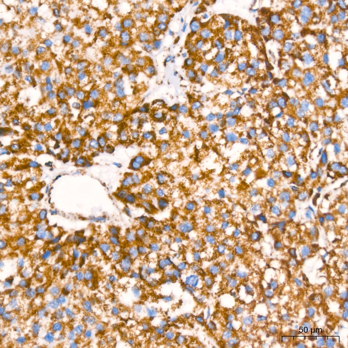
Immunohistochemistry analysis of paraffin-embedded Human liver cancer using NDUFB8 Rabbit mAb (CAB19732) at dilution of 1:200 (40x lens). High pressure antigen retrieval performed with 0.01M Citrate buffer (pH 6.0) prior to IHC staining.
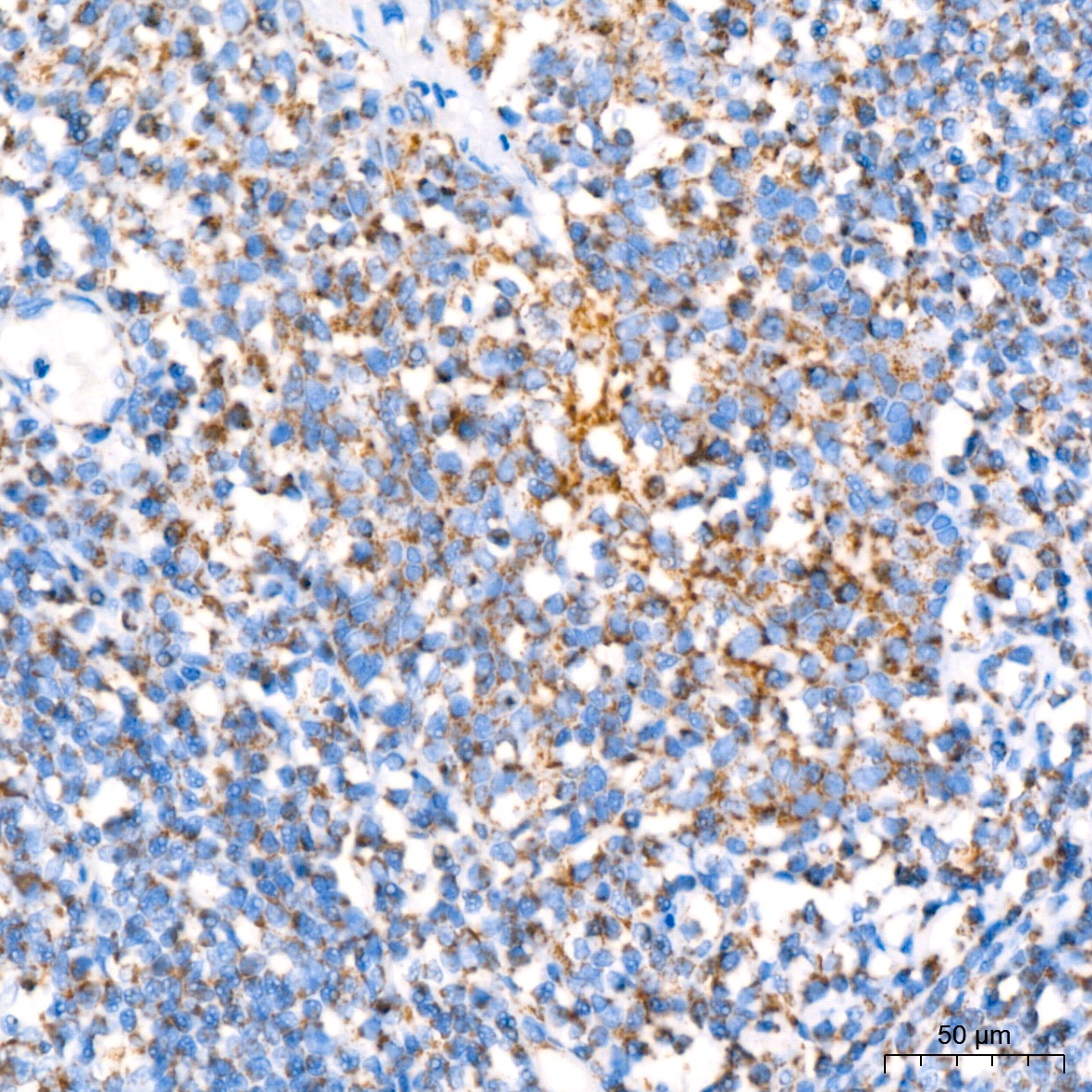
Immunohistochemistry analysis of paraffin-embedded Human tonsil using NDUFB8 Rabbit mAb (CAB19732) at dilution of 1:200 (40x lens). High pressure antigen retrieval performed with 0.01M Citrate buffer (pH 6.0) prior to IHC staining.
Immunohistochemistry analysis of paraffin-embedded Rat kidney using NDUFB8 Rabbit mAb (CAB19732) at dilution of 1:200 (40x lens). High pressure antigen retrieval performed with 0.01M Citrate buffer (pH 6.0) prior to IHC staining.
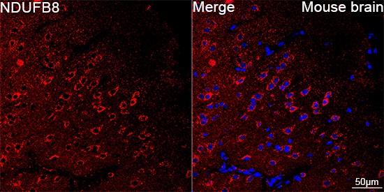
Confocal imaging of paraffin-embedded Mouse brain using NDUFB8 Rabbit mAb (CAB19732, dilution 1:200) followed by a further incubation with Cy3 Goat Anti-Rabbit IgG (H+L) (CABS007, dilution 1:500) (Red). DAPI was used for nuclear staining (Blue). Objective: 40x. Perform microwave antigen retrieval with 0.01 M citrate buffer (pH 6.0) prior to IF staining.


![Anti-NDUFB8 [R03-8G2] Monoclonal Antibody (AGMB01869)](https://cdn11.bigcommerce.com/s-h68l9z2lnx/images/stencil/590x590/products/273158/676798/anti-ndufb8-r03-8g2-monoclonal-antibody-agmb01869__74000.1773030613.jpg?c=2 "Anti-NDUFB8 [R03-8G2] Monoclonal Antibody (AGMB01869)")


