The NRF2 Antibody (CAB1244) is a high-quality antibody developed for reliable detection and analysis of target proteins. The antibody, raised in rabbits, is highly reactive with human samples and has been validated for use in various applications, including Western blot and immunohistochemistry. By binding to NFE2L2 protein, this antibody enables accurate detection and analysis in multiple cell types, making it an essential tool for studies in oxidative stress, inflammation, and cancer research.NFE2L2, also known as nuclear factor erythroid 2-related factor 2, is a crucial regulator of cellular defense mechanisms against oxidative stress and inflammation. Its involvement in the activation of antioxidant and detoxification genes makes it a central player in maintaining cellular homeostasis and protecting cells from damage.
This antibody is validated for use in WB, IHC-P, ELISA applications and has demonstrated reactivity against Human, Mouse, Rat samples.
Product Name:
NRF2 Antibody
SKU:
CAB1244
Size:
20μL, 100μL
Reactivity:
Human, Mouse, Rat
Conjugate:
Unconjugated
Immunogen:
Recombinant protein (or fragment).This information is considered to be commercially sensitive.
Recommended starting concentration is 1 μg/mL. Please optimize the concentration based on your specific assay requirements.
Synonyms:
NRF2, HEBP1, Nrf-2, IMDDHH
Positive Sample:
NIH/3T3 treated with MG132, C6 treated with MG132
Cellular Localization:
Cytoplasm, Nucleus, Cytosol.
Calculated MW:
68kDa
Observed MW:
100kDa
This gene encodes a transcription factor which is a member of a small family of basic leucine zipper (bZIP) proteins. The encoded transcription factor regulates genes which contain antioxidant response elements (ARE) in their promoters; many of these genes encode proteins involved in response to injury and inflammation which includes the production of free radicals. Multiple transcript variants encoding different isoforms have been characterized for this gene.
Purification Method
Affinity purification
Gene ID
4780
RRID
AB_2759282
Buffer Information
Store at -20℃. Avoid freeze / thaw cycles. Buffer: PBS containing 50% glycerol, preserved with proclin300 or sodium azide, pH 7.3.
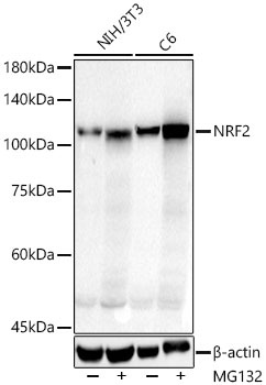
Western blot analysis of lysates from NIH/3T3,C6 cells using NRF2 Rabbit pAb (CAB1244) at 1:1000 dilution. NIH/3T3 cells and C6 cells were treated with MG132(10 μM) at 37℃ for 90 minutes. Secondary antibody: HRP-conjugated Goat anti-Rabbit IgG (H+L) (CABS014) at 1:10000 dilution. Lysates/proteins: 45 μg per lane. Blocking buffer: 3% nonfat dry milk in TBST. Detection: ECL Basic Kit (AbGn00020). Exposure time: 10s.
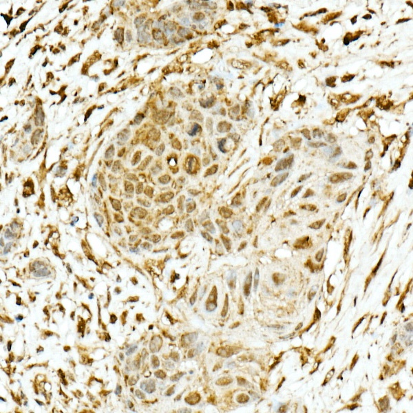
Immunohistochemistry analysis of paraffin-embedded Human esophageal cancer using NRF2 Rabbit pAb (CAB1244) at dilution of 1:200 (40x lens). High pressure antigen retrieval performed with 0.01M Citrate buffer (pH 6.0) prior to IHC staining.
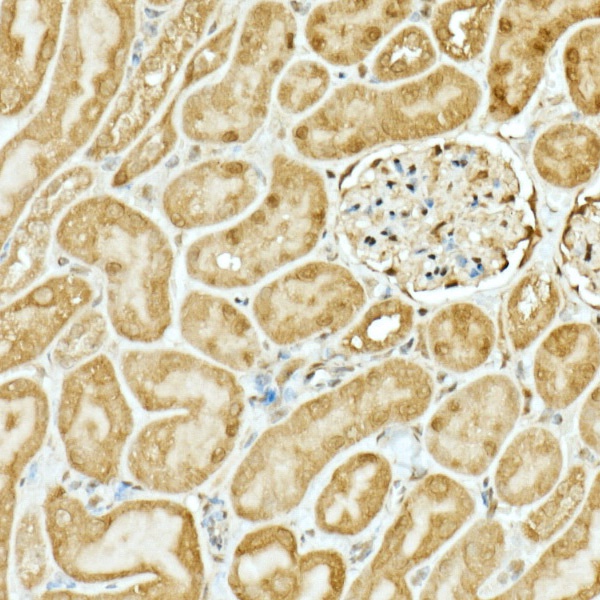
Immunohistochemistry analysis of paraffin-embedded Mouse kidney using NRF2 Rabbit pAb (CAB1244) at dilution of 1:200 (40x lens). High pressure antigen retrieval performed with 0.01M Citrate buffer (pH 6.0) prior to IHC staining.
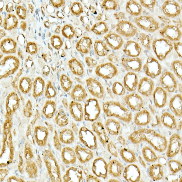
Immunohistochemistry analysis of paraffin-embedded Rat kidney using NRF2 Rabbit pAb (CAB1244) at dilution of 1:200 (40x lens). High pressure antigen retrieval performed with 0.01M Citrate buffer (pH 6.0) prior to IHC staining.