The NPC2 Antibody (CAB5413) is a high-quality antibody developed for reliable detection and analysis of target proteins. This antibody, raised in rabbits, is highly specific for human samples and is validated for use in various applications, including Western blot and immunofluorescence.NPC2 plays a crucial role in intracellular cholesterol trafficking and mutations in the NPC2 gene have been linked to Niemann-Pick disease type C2, a rare lysosomal storage disorder. By using the NPC2 Polyclonal Antibody, researchers can study the expression and localization of NPC2 in different cell types and tissues, shedding light on its function in cholesterol metabolism and potential implications in disease.
This antibody is validated for use in WB, IHC-P, ELISA, IF-P applications and has demonstrated reactivity against Human, Mouse, Rat samples.
Product Name:
NPC2 Antibody
SKU:
CAB5413
Size:
20μL, 100μL
Reactivity:
Human, Mouse, Rat
Conjugate:
Unconjugated
Immunogen:
Recombinant protein (or fragment).This information is considered to be commercially sensitive.
Recommended starting concentration is 1 μg/mL. Please optimize the concentration based on your specific assay requirements.
Synonyms:
HE1, EDDM1, NPC2
Positive Sample:
Mouse lung, 293T, NIH/3T3, Hep G2
Cellular Localization:
Endoplasmic Reticulum, Lysosome, Secreted.
Calculated MW:
17kDa
Observed MW:
19kDa/17-21kDa
This gene encodes a protein containing a lipid recognition domain. The encoded protein may function in regulating the transport of cholesterol through the late endosomal/lysosomal system. Mutations in this gene have been associated with Niemann-Pick disease, type C2 and frontal lobe atrophy.
Purification Method
Affinity purification
Gene ID
10577
RRID
AB_2766221
Buffer Information
Store at -20℃. Avoid freeze / thaw cycles. Buffer: PBS containing 50% glycerol, preserved with proclin300 or sodium azide, pH 7.3.
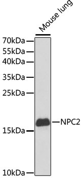
Western blot analysis of lysates from mouse lung, using NPC2 Rabbit pAb (CAB5413) at 1:1000 dilution. Secondary antibody: HRP-conjugated Goat anti-Rabbit IgG (H+L) (CABS014) at 1:10000 dilution. Lysates/proteins: 25μg per lane. Blocking buffer: 3% nonfat dry milk in TBST. Detection: ECL Basic Kit (AbGn00020). Exposure time: 90s.
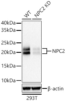
Western blot analysis of lysates from wild type (WT) and NPC2 knockdown (KD) 293T cells using NPC2 Rabbit pAb (CAB5413) at 1:1000 dilution incubated overnight at 4℃. Secondary antibody: HRP-conjugated Goat anti-Rabbit IgG (H+L) (CABS014) at 1:10000 dilution. Lysates/proteins: 25 μg per lane. Blocking buffer: 3% nonfat dry milk in TBST. Detection: ECL Basic Kit (AbGn00020). Exposure time: 45s.
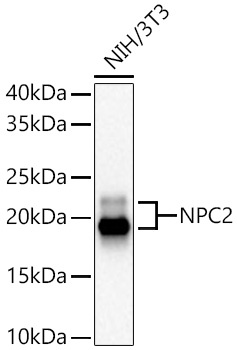
Western blot analysis of lysates from NIH/3T3 cells using NPC2 Rabbit pAb (CAB5413) at 1:1000 dilution incubated overnight at 4℃. Secondary antibody: HRP-conjugated Goat anti-Rabbit IgG (H+L) (CABS014) at 1:10000 dilution. Lysates/proteins: 25 μg per lane. Blocking buffer: 3% nonfat dry milk in TBST. Detection: ECL Basic Kit (AbGn00020). Exposure time: 30s.
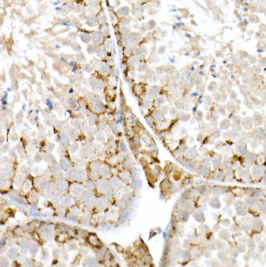
Immunohistochemistry analysis of paraffin-embedded Mouse testis using NPC2 Rabbit pAb (CAB5413) at dilution of 1:50 (40x lens). High pressure antigen retrieval performed with 0.01M Citrate buffer (pH 6.0) prior to IHC staining.
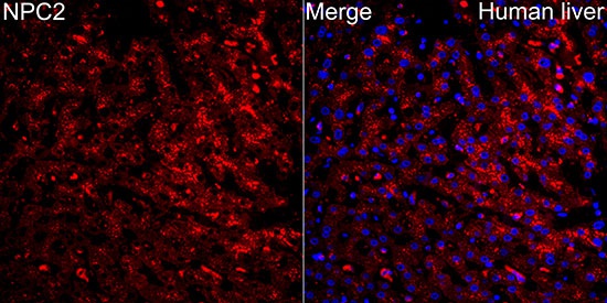
Immunofluorescence analysis of Human liver tissue using NPC2 Rabbit pAb (CAB5413) at a dilution of 1:100 (40x lens). Secondary antibody: Cy3-conjugated Goat anti-Rabbit IgG (H+L)(CABS007) at 1:500 dilution. Blue: DAPI for nuclear staining. High pressure antigen retrieval performed with 0.01M Citrate Buffer (pH 6.0) prior to IF staining.
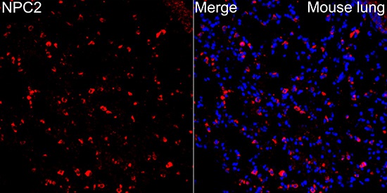
Immunofluorescence analysis of Mouse lung tissue using NPC2 Rabbit pAb (CAB5413) at a dilution of 1:100 (40x lens). Secondary antibody: Cy3-conjugated Goat anti-Rabbit IgG (H+L)(CABS007) at 1:500 dilution. Blue: DAPI for nuclear staining. High pressure antigen retrieval performed with 0.01M Citrate Buffer (pH 6.0) prior to IF staining.