The PDE1A Antibody (CAB10457) is a high-quality antibody developed for reliable detection and analysis of target proteins. This antibody, produced through rabbit immunization, exhibits strong reactivity with human samples and has been validated for use in Western blot applications.PDE1A is known to be involved in various physiological processes, including smooth muscle contraction, cardiac function, and neuronal signaling. Dysregulation of PDE1A has been linked to conditions such as heart failure, hypertension, and neurological disorders.
This antibody is validated for use in WB, ELISA applications and has demonstrated reactivity against Mouse, Rat samples.
Product Name:
PDE1A Antibody
SKU:
CAB10457
Size:
20μL, 100μL
Reactivity:
Mouse, Rat
Conjugate:
Unconjugated
Immunogen:
Recombinant protein (or fragment).This information is considered to be commercially sensitive.
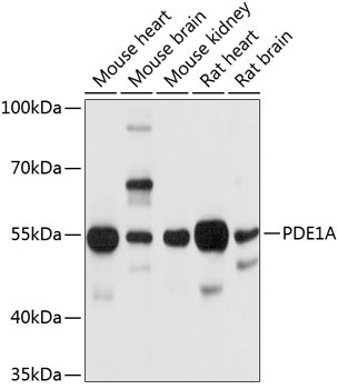
Mouse heart, Mouse brain, Mouse kidney, Rat heart, Rat brain
Cellular Localization:
Cytosol.
Calculated MW:
61kDa
Observed MW:
55kDa
Cyclic nucleotide phosphodiesterases (PDEs) play a role in signal transduction by regulating intracellular cyclic nucleotide concentrations through hydrolysis of cAMP and/or cGMP to their respective nucleoside 5-prime monophosphates. Members of the PDE1 family, such as PDE1A, are Ca(2+)/calmodulin (see CALM1; MIM 114180)-dependent PDEs (CaM-PDEs) that are activated by calmodulin in the presence of Ca(2+) (Michibata et al., 2001 [PubMed 11342109]; Fidock et al., 2002 [PubMed 11747989]).
Purification Method
Affinity purification
Gene ID
5136
RRID
AB_2758005
Buffer Information
Store at -20℃. Avoid freeze / thaw cycles. Buffer: PBS containing 50% glycerol, preserved with proclin300 or sodium azide, pH 7.3.
Western blot analysis of various lysates using PDE1A Rabbit pAb (CAB10457) at 1:1000 dilution. Secondary antibody: HRP-conjugated Goat anti-Rabbit IgG (H+L) (CABS014) at 1:10000 dilution. Lysates/proteins: 25μg per lane. Blocking buffer: 3% nonfat dry milk in TBST. Detection: ECL Basic Kit (AbGn00020). Exposure time: 10s.