The PDE6B Antibody (CAB6942) is a high-quality antibody developed for reliable detection and analysis of target proteins. Raised in rabbits, this antibody is highly specific to human PDE6B samples and is validated for use in Western blot applications. By binding to the PDE6B protein, this antibody enables detection and analysis of PDE6B in various cell types, making it ideal for studies in vision research and retinal diseases.
This antibody is validated for use in WB, IHC-P, ELISA applications and has demonstrated reactivity against Human, Mouse, Rat samples.
Product Name:
PDE6B Antibody
SKU:
CAB6942
Size:
20μL, 100μL
Reactivity:
Human, Mouse, Rat
Conjugate:
Unconjugated
Immunogen:
Recombinant protein (or fragment).This information is considered to be commercially sensitive.
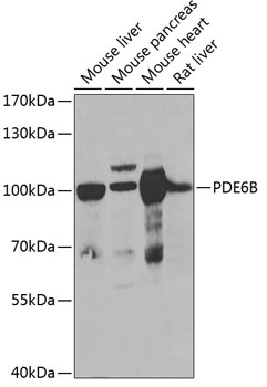
Mouse liver, Mouse pancreas, Mouse heart, Rat liver
Cellular Localization:
Lipid-Anchor, Membrane.
Calculated MW:
98kDa
Observed MW:
98kDa
Photon absorption triggers a signaling cascade in rod photoreceptors that activates cGMP phosphodiesterase (PDE), resulting in the rapid hydrolysis of cGMP, closure of cGMP-gated cation channels, and hyperpolarization of the cell. PDE is a peripheral membrane heterotrimeric enzyme made up of alpha, beta, and gamma subunits. This gene encodes the beta subunit. Mutations in this gene result in retinitis pigmentosa and autosomal dominant congenital stationary night blindness. Multiple transcript variants encoding different isoforms have been found for this gene.
Purification Method
Affinity purification
Gene ID
5158
RRID
AB_2767500
Buffer Information
Store at -20℃. Avoid freeze / thaw cycles. Buffer: PBS containing 50% glycerol, preserved with proclin300 or sodium azide, pH 7.3.
Western blot analysis of various lysates using PDE6B Rabbit pAb (CAB6942) at 1:1000 dilution. Secondary antibody: HRP-conjugated Goat anti-Rabbit IgG (H+L) (CABS014) at 1:10000 dilution. Lysates/proteins: 25μg per lane. Blocking buffer: 3% nonfat dry milk in TBST. Detection: ECL Basic Kit (AbGn00020). Exposure time: 90s.
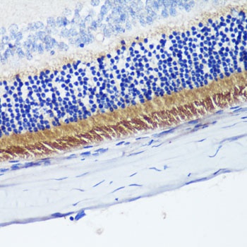
Immunohistochemistry analysis of paraffin-embedded Rat retina using PDE6B Rabbit pAb (CAB6942) at dilution of 1:200 (40x lens). Microwave antigen retrieval performed with 0.01M PBS Buffer (pH 7.2) prior to IHC staining.