The PDE6G Antibody (CAB17516) is a high-quality antibody developed for reliable detection and analysis of target proteins. This antibody, raised in rabbits, demonstrates high reactivity with human samples and has been validated for use in Western blot applications. By binding to the PDE6G protein, this antibody enables accurate detection and analysis in a variety of cell types, making it an ideal tool for studies in vision research and retinal disorders.PDE6G is a crucial component of the phototransduction cascade in rod cells, where it serves to regulate cGMP levels and facilitate the visual response to light.
This antibody is validated for use in WB, IF/ICC, ELISA applications and has demonstrated reactivity against Human, Mouse, Rat samples.
Product Name:
PDE6G Antibody
SKU:
CAB17516
Size:
20μL, 100μL
Reactivity:
Human, Mouse, Rat
Conjugate:
Unconjugated
Immunogen:
Recombinant protein (or fragment).This information is considered to be commercially sensitive.
This gene encodes the gamma subunit of cyclic GMP-phosphodiesterase, which is composed of alpha- and beta- catalytic subunits and two identical, inhibitory gamma subunits. This gene is expressed in rod photoreceptors and functions in the phototransduction signaling cascade. It is also expressed in a variety of other tissues, and has been shown to regulate the c-Src protein kinase and G-protein-coupled receptor kinase 2. Alternative splicing results in multiple transcript variants.
Purification Method
Affinity purification
Gene ID
5148
RRID
AB_2770829
Buffer Information
Store at -20℃. Avoid freeze / thaw cycles. Buffer: PBS with 0.01% thimerosal,50% glycerol,pH7.3.
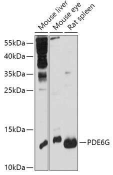
Western blot analysis of various lysates using PDE6G Rabbit pAb (CAB17516) at 1:1000 dilution. Secondary antibody: HRP-conjugated Goat anti-Rabbit IgG (H+L) (CABS014) at 1:10000 dilution. Lysates/proteins: 25μg per lane. Blocking buffer: 3% nonfat dry milk in TBST. Detection: ECL Basic Kit (AbGn00020). Exposure time: 30s.
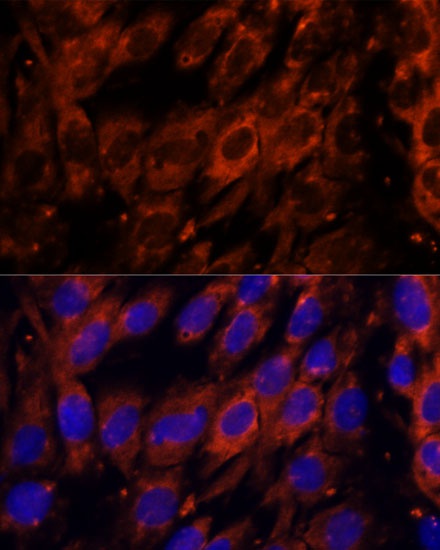
Immunofluorescence analysis of C6 cells using PDE6G Rabbit pAb (CAB17516) at dilution of 1:100. Secondary antibody: Cy3-conjugated Goat anti-Rabbit IgG (H+L) (CABS007) at 1:500 dilution. Blue: DAPI for nuclear staining.
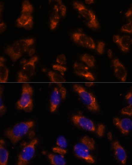
Immunofluorescence analysis of U-2 OS cells using PDE6G Rabbit pAb (CAB17516) at dilution of 1:100. Secondary antibody: Cy3-conjugated Goat anti-Rabbit IgG (H+L) (CABS007) at 1:500 dilution. Blue: DAPI for nuclear staining.