The PDHA2 Antibody (CAB14994) is a high-quality antibody developed for reliable detection and analysis of target proteins. This antibody, produced in rabbits, is specifically designed for use in Western blot applications and is highly reactive with human samples.PDHA2 is an essential component of the pyruvate dehydrogenase complex, which plays a crucial role in the conversion of pyruvate to acetyl-CoA in the mitochondria. Dysregulation of this complex has been linked to various metabolic disorders and diseases, making PDHA2 a key target for research in the fields of metabolism and biochemistry.
This antibody is validated for use in WB, IF/ICC, ELISA applications and has demonstrated reactivity against Human, Mouse, Rat samples.
Product Name:
PDHA2 Antibody
SKU:
CAB14994
Size:
20μL, 100μL
Reactivity:
Human, Mouse, Rat
Conjugate:
Unconjugated
Immunogen:
Recombinant protein (or fragment).This information is considered to be commercially sensitive.
Recommended starting concentration is 1 μg/mL. Please optimize the concentration based on your specific assay requirements.
Synonyms:
Pdhal, PDHA2
Positive Sample:
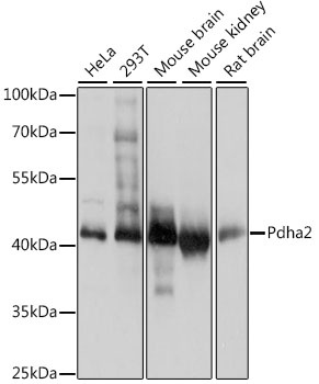
HeLa, 293T, Mouse brain, Mouse kidney, Rat brain
Cellular Localization:
Mitochondrion Matrix.
Calculated MW:
43kDa
Observed MW:
43kDa
Predicted to enable pyruvate dehydrogenase (acetyl-transferring) activity. Predicted to contribute to pyruvate dehydrogenase (NAD+) activity. Predicted to be involved in acetyl-CoA biosynthetic process from pyruvate. Predicted to act upstream of or within mitochondrial acetyl-CoA biosynthetic process from pyruvate. Located in mitochondrion. Is expressed in nervous system and testis. Orthologous to human PDHA2 (pyruvate dehydrogenase E1 subunit alpha 2).
Purification Method
Affinity purification
Gene ID
18598
RRID
AB_2761877
Buffer Information
Store at -20℃. Avoid freeze / thaw cycles. Buffer: PBS with 0.01% thimerosal,50% glycerol,pH7.3.
Western blot analysis of various lysates using PDHA2 Rabbit pAb (CAB14994) at 1:1000 dilution. Secondary antibody: HRP-conjugated Goat anti-Rabbit IgG (H+L) (CABS014) at 1:10000 dilution. Lysates/proteins: 25μg per lane. Blocking buffer: 3% nonfat dry milk in TBST. Detection: ECL Basic Kit (AbGn00020). Exposure time: 1s.
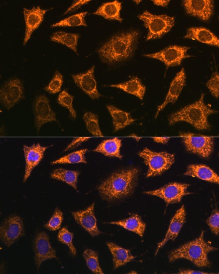
Immunofluorescence analysis of L929 cells using PDHA2 Rabbit pAb (CAB14994) at dilution of 1:100 (40x lens). Secondary antibody: Cy3-conjugated Goat anti-Rabbit IgG (H+L) (CABS007) at 1:500 dilution. Blue: DAPI for nuclear staining.