The PPL Antibody (CAB6950) is a high-quality antibody developed for reliable detection and analysis of target proteins. This antibody, generated in rabbits, is highly specific for human samples and has been validated for use in Western blot applications. By targeting the periplakin protein, researchers can analyze its expression and localization in various cell types, providing valuable insights into cell adhesion mechanisms and potential implications in diseases such as cancer and skin disorders.
This antibody is validated for use in WB, IF/ICC, ELISA applications and has demonstrated reactivity against Human, Mouse, Rat samples.
Product Name:
PPL Antibody
SKU:
CAB6950
Size:
20μL, 100μL
Reactivity:
Human, Mouse, Rat
Conjugate:
Unconjugated
Immunogen:
Recombinant protein (or fragment).This information is considered to be commercially sensitive.
The protein encoded by this gene is a component of desmosomes and of the epidermal cornified envelope in keratinocytes. The N-terminal domain of this protein interacts with the plasma membrane and its C-terminus interacts with intermediate filaments. Through its rod domain, this protein forms complexes with envoplakin. This protein may serve as a link between the cornified envelope and desmosomes as well as intermediate filaments. AKT1/PKB, a protein kinase mediating a variety of cell growth and survival signaling processes, is reported to interact with this protein, suggesting a possible role for this protein as a localization signal in AKT1-mediated signaling.
Purification Method
Affinity purification
Gene ID
5493
RRID
AB_2767508
Buffer Information
Store at -20℃. Avoid freeze / thaw cycles. Buffer: PBS containing 50% glycerol, preserved with proclin300 or sodium azide, pH 7.3.
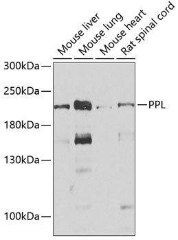
Western blot analysis of various lysates using PPL Rabbit pAb (CAB6950) at 1:1000 dilution. Secondary antibody: HRP-conjugated Goat anti-Rabbit IgG (H+L) (CABS014) at 1:10000 dilution. Lysates/proteins: 25μg per lane. Blocking buffer: 3% nonfat dry milk in TBST. Detection: ECL Enhanced Kit (AbGn00021). Exposure time: 30s.
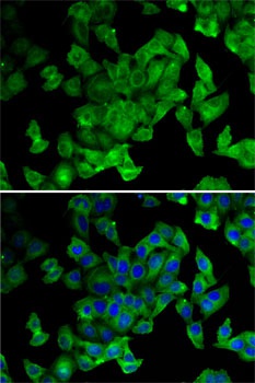
Immunofluorescence analysis of MCF7 cells using PPL Rabbit pAb (CAB6950). Secondary antibody: Cy3-conjugated Goat anti-Rabbit IgG (H+L) (CABS007) at 1:500 dilution. Blue: DAPI for nuclear staining.