The Phospho-CFL1-S3 Antibody (CABP0178) is a high-quality antibody developed for reliable detection and analysis of target proteins. This antibody, generated in rabbits, exhibits high specificity for human samples and has been validated for use in Western blot and immunohistochemistry applications.Cofilin-1 is a key regulator of actin cytoskeleton dynamics, playing a vital role in processes such as cell migration, adhesion, and invasion. Phosphorylation of Cofilin-1 at Serine 3 modulates its activity, influencing actin filament assembly/disassembly and cellular morphology.
This antibody is validated for use in WB, IHC-P, IF/ICC, ELISA applications and has demonstrated reactivity against Human, Mouse, Rat samples.
Product Name:
Phospho-CFL1-S3 Antibody
SKU:
CABP0178
Size:
20μL, 100μL
Reactivity:
Human, Mouse, Rat
Conjugate:
Unconjugated
Immunogen:
Synthetic peptide. This information is considered to be commercially sensitive.
Sequence:
MASG V
Tested Applications:
WBIHC-PIF/ICCELISA
Recommended Dilution:
WB
1:500 - 1:2000
IHC-P
1:50 - 1:100
IF/ICC
1:100 - 1:200
ELISA
Recommended starting concentration is 1 μg/mL. Please optimize the concentration based on your specific assay requirements.
The protein encoded by this gene can polymerize and depolymerize F-actin and G-actin in a pH-dependent manner. Increased phosphorylation of this protein by LIM kinase aids in Rho-induced reorganization of the actin cytoskeleton. Cofilin is a widely distributed intracellular actin-modulating protein that binds and depolymerizes filamentous F-actin and inhibits the polymerization of monomeric G-actin in a pH-dependent manner. It is involved in the translocation of actin-cofilin complex from cytoplasm to nucleus.
Purification Method
Affinity purification
Gene ID
1072
RRID
AB_2770988
Buffer Information
Store at -20℃. Avoid freeze / thaw cycles. Buffer: PBS with 0.09% Sodium azide,50% glycerol,pH7.3.
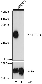
Western blot analysis of lysates from NIH/3T3 cells, using Phospho-CFL1-S3 Rabbit pAb (CAB1704). NIH/3T3 cells were treated by CIP(20uL/400ul) at 37℃ for 1 hour. Secondary antibody: HRP-conjugated Goat anti-Rabbit IgG (H+L) (CABS014) at 1:10000 dilution. Lysates/proteins: 25μg per lane. Blocking buffer: 3% BSA. Detection: ECL Basic Kit (AbGn00020). Exposure time: 3s.
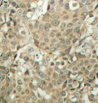
Immunohistochemistry analysis of paraffin-embedded Human breast carcinoma tissue, using Phospho-CFL1-S3 Rabbit pAb (CABP0178). Microwave antigen retrieval performed with 0.01M Tris/EDTA Buffer (pH 9.0) prior to IHC staining.