The Phospho-GSK3beta-S9 Monoclonal Antibody (CABP1088) is a high-quality antibody developed for reliable detection and analysis of target proteins. This antibody, generated in rabbits, specifically recognizes the phosphorylated form of GSK3beta at serine 9 and is validated for use in Western blot and immunohistochemistry assays.GSK3beta is a critical regulator of cell growth, proliferation, and survival, and its activity is tightly controlled by phosphorylation events. Phosphorylation of GSK3beta at serine 9 inhibits its activity, leading to downstream effects on cell signaling pathways.
This antibody is validated for use in WB, IHC-P, ELISA applications and has demonstrated reactivity against Human samples.
Product Name:
Phospho-GSK3beta-S9 Monoclonal Antibody
SKU:
CABP1088
Size:
20μL, 100μL
Reactivity:
Human
Clone Number:
ARC0069
Conjugate:
Unconjugated
Immunogen:
Synthetic peptide. This information is considered to be commercially sensitive.
Sequence:
Email for sequence
Tested Applications:
WBIHC-PELISA
Recommended Dilution:
WB
1:1000 - 1:6000
IHC-P
1:50 - 1:200
ELISA
Recommended starting concentration is 1 μg/mL. Please optimize the concentration based on your specific assay requirements.
Synonyms:
GSK3B, gsk-3β, Phospho-GSK3β-S9
Positive Sample:
HeLa treated with Calyculin A
Cellular Localization:
Cell Membrane, Cytoplasm, Nucleus.
Calculated MW:
47kDa
Observed MW:
47kDa
The protein encoded by this gene is a serine-threonine kinase belonging to the glycogen synthase kinase subfamily. It is a negative regulator of glucose homeostasis and is involved in energy metabolism, inflammation, ER-stress, mitochondrial dysfunction, and apoptotic pathways. Defects in this gene have been associated with Parkinson disease and Alzheimer disease.
Purification Method
Affinity purification
Gene ID
2932
RRID
AB_2863959
Buffer Information
Store at -20℃. Avoid freeze / thaw cycles. Buffer: PBS containing 50% glycerol and 0.05% BSA, preserved with proclin300 or sodium azide, pH 7.3.
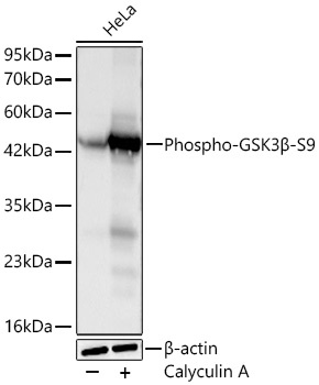
Western blot analysis of lysates from HeLa cells using Phospho-GSK3β-S9 Rabbit mAb (CABP1088) at 1:3000 dilution incubated at room temperature for 1.5 hours. HeLa cells were treated with Calyculin A (100 nM) at 37℃ for 30 minutes after serum-starvation overnight. Secondary antibody: HRP-conjugated Goat anti-Rabbit IgG (H+L) (CABS014) at 1:10000 dilution. Lysates/proteins: 30 μg per lane. Blocking buffer: 3% nonfat dry milk in TBST. Detection: ECL Basic Kit (AbGn00020). Exposure time: 20s.
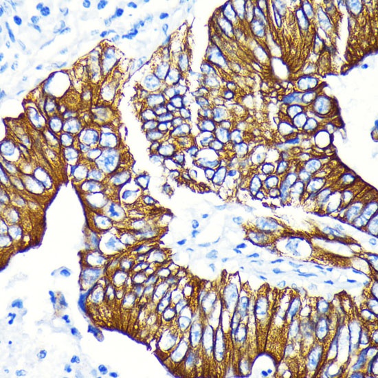
Immunohistochemistry analysis of paraffin-embedded Human colon carcinoma tissue using Phospho-GSK3β-S9 Rabbit mAb (CABP1088) at dilution of 1:100 (40x lens). Microwave antigen retrieval performed with 0.01M Tris-EDTA Buffer (pH 9.0) prior to IHC staining.
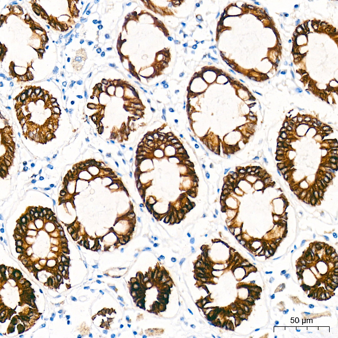
Immunohistochemistry analysis of paraffin-embedded Human colon tissue using Phospho-GSK3β-S9 Rabbit mAb (CABP1088) at a dilution of 1:200 (40x lens). High pressure antigen retrieval performed with 0.01M Citrate Buffer (pH 6.0) prior to IHC staining.