The Phospho-PDHA1-S293 Monoclonal Antibody (CABP1022) is a high-quality antibody developed for reliable detection and analysis of target proteins. This antibody, generated in rabbits, exhibits high specificity for human samples and has been validated for use in Western blot applications.PDHA1 phosphorylation at serine 293 plays a crucial role in the regulation of its enzymatic activity, impacting cellular energy metabolism and oxidative stress response. The Phospho-PDHA1 (S293) antibody allows for the detection and quantification of phosphorylated PDHA1, providing valuable insights into the metabolic processes and signaling pathways involved in cell function.
This antibody is validated for use in WB, IP, ELISA applications and has demonstrated reactivity against Human, Mouse, Rat samples.
Product Name:
Phospho-PDHA1-S293 Monoclonal Antibody
SKU:
CABP1022
Size:
20μL, 100μL
Reactivity:
Human, Mouse, Rat
Clone Number:
ARC53489
Conjugate:
Unconjugated
Immunogen:
Synthetic peptide. This information is considered to be commercially sensitive.
Sequence:
GHSM SD
Tested Applications:
WBIPELISA
Recommended Dilution:
WB
1:5000 - 1:40000
IP
0.5μg-4μg antibody for 200μg-400μg extracts of whole cells
ELISA
Recommended starting concentration is 1 μg/mL. Please optimize the concentration based on your specific assay requirements.
The pyruvate dehydrogenase (PDH) complex is a nuclear-encoded mitochondrial multienzyme complex that catalyzes the overall conversion of pyruvate to acetyl-CoA and CO(2), and provides the primary link between glycolysis and the tricarboxylic acid (TCA) cycle. The PDH complex is composed of multiple copies of three enzymatic components: pyruvate dehydrogenase (E1), dihydrolipoamide acetyltransferase (E2) and lipoamide dehydrogenase (E3). The E1 enzyme is a heterotetramer of two alpha and two beta subunits. This gene encodes the E1 alpha 1 subunit containing the E1 active site, and plays a key role in the function of the PDH complex. Mutations in this gene are associated with pyruvate dehydrogenase E1-alpha deficiency and X-linked Leigh syndrome. Alternatively spliced transcript variants encoding different isoforms have been found for this gene.
Purification Method
Affinity purification
Gene ID
5160
RRID
AB_2863907
Buffer Information
Store at -20℃. Avoid freeze / thaw cycles. Buffer: PBS containing 50% glycerol and 0.05% BSA, preserved with proclin300 or sodium azide, pH 7.3.
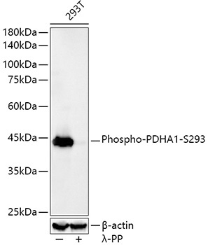
Western blot analysis of lysates from HeLa cells, using Phospho-PDHA1-S293 Rabbit mAb (CABP1022) at 1:23000 dilution. 293T cells were treated by λ-PP mixed solution (1ul) at 30℃ for 30 minutes. Secondary antibody: HRP-conjugated Goat anti-Rabbit IgG (H+L) (CABS014) at 1:10000 dilution. Lysates/proteins: 25μg per lane. Blocking buffer: 3% nonfat dry milk in TBST. Detection: ECL Basic Kit (AbGn00020). Exposure time: 30s.
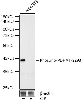
Western blot analysis of lysates from NIH/3T3 cells, using Phospho-PDHA1-S293 Rabbit mAb (CABP1022) at1:23000 dilution. NIH/3T3 cells were treated by CIP(20uL/400ul) at 37℃ for 1 hour. Secondary antibody: HRP-conjugated Goat anti-Rabbit IgG (H+L) (CABS014) at 1:10000 dilution. Lysates/proteins: 25μg per lane. Blocking buffer: 3% nonfat dry milk in TBST. Detection: ECL Basic Kit (AbGn00020). Exposure time: 30s.