The Phospho-p90Rsk/RSK1/RPS6KA1-S380 Monoclonal Antibody (CABP1147) is a high-quality antibody developed for reliable detection and analysis of target proteins. RSK1 is a kinase involved in cell proliferation, differentiation, and survival, making it a key player in cancer research and signaling pathways.This antibody, generated in rabbits, is highly specific and sensitive towards human samples, making it ideal for Western blot applications. It binds specifically to the phosphorylated form of RSK1 at serine 380, allowing for precise detection and analysis in various cell types.
This antibody is validated for use in WB, ELISA applications and has demonstrated reactivity against Human, Mouse, Rat samples.
A-431 treated with EGF, NIH/3T3 treated with PDGF, C6 treated with PMA
Cellular Localization:
Cytoplasm, Nucleus.
Calculated MW:
83kDa
Observed MW:
90kDa
This gene encodes a member of the RSK (ribosomal S6 kinase) family of serine/threonine kinases. This kinase contains 2 nonidentical kinase catalytic domains and phosphorylates various substrates, including members of the mitogen-activated kinase (MAPK) signalling pathway. The activity of this protein has been implicated in controlling cell growth and differentiation. Alternate transcriptional splice variants, encoding different isoforms, have been characterized.
Purification Method
Affinity purification
Gene ID
6195
RRID
AB_2864012
Buffer Information
Store at -20℃. Avoid freeze / thaw cycles. Buffer: PBS containing 50% glycerol and 0.05% BSA, preserved with proclin300 or sodium azide, pH 7.3.
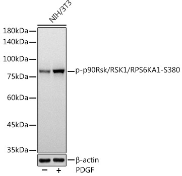
Western blot analysis of lysates from NIH/3T3 cells, using Phospho-p90Rsk/RSK1/RPS6KA1-S380 Rabbit mAb (CABP1147) at 1:1000 dilution. NIH/3T3 cells were treated with PDGF (100 ng/ml) at 37℃ for 30 minutes after serum-starvation overnight. Secondary antibody: HRP-conjugated Goat anti-Rabbit IgG (H+L) (CABS014) at 1:10000 dilution. Lysates/proteins: 25μg per lane. Blocking buffer: 3% nonfat dry milk in TBST. Detection: ECL Basic Kit (AbGn00020). Exposure time: 3s.
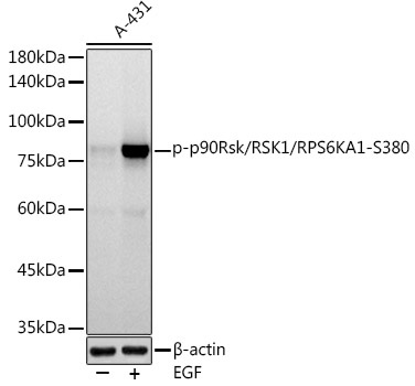
Western blot analysis of lysates from A-431 cells, using Phospho-p90Rsk/RSK1/RPS6KA1-S380 Rabbit mAb (CABP1147) at 1:1000 dilution. A-431 cells were treated with EGF (100 ng/ml) at 37℃ for 30 minutes after serum-starvation overnight. Secondary antibody: HRP-conjugated Goat anti-Rabbit IgG (H+L) (CABS014) at 1:10000 dilution. Lysates/proteins: 25μg per lane. Blocking buffer: 3% nonfat dry milk in TBST. Detection: ECL Basic Kit (AbGn00020). Exposure time: 90s.


![Anti-Phospho-RSK1 p90 (Ser380) [R05-7F3] Monoclonal Antibody (AGMB05241)](https://cdn11.bigcommerce.com/s-h68l9z2lnx/images/stencil/590x590/products/276526/676431/anti-phospho-rsk1-p90-ser380-r05-7f3-monoclonal-antibody-agmb05241__04257.1773029421.jpg?c=2 "Anti-Phospho-RSK1 p90 (Ser380) [R05-7F3] Monoclonal Antibody (AGMB05241)")
![Anti-Phospho-RSK1 p90 (Thr359/Ser363) [R01-5B1] Monoclonal Antibody (AGMB05061)](https://cdn11.bigcommerce.com/s-h68l9z2lnx/images/stencil/590x590/products/276346/677018/anti-phospho-rsk1-p90-thr359ser363-r01-5b1-monoclonal-antibody-agmb05061__45648.1773031236.jpg?c=2 "Anti-Phospho-RSK1 p90 (Thr359/Ser363) [R01-5B1] Monoclonal Antibody (AGMB05061)")
![Anti-Phospho-RSK1 p90 (Ser380) [R03-8I3] Monoclonal Antibody (AGMB05062)](https://cdn11.bigcommerce.com/s-h68l9z2lnx/images/stencil/590x590/products/276347/679160/anti-phospho-rsk1-p90-ser380-r03-8i3-monoclonal-antibody-agmb05062__68365.1773038058.jpg?c=2 "Anti-Phospho-RSK1 p90 (Ser380) [R03-8I3] Monoclonal Antibody (AGMB05062)")
![Anti-Phospho-RSK1 p90 (Thr359/Ser363) [R33-5H-9] Monoclonal Antibody (AGMB05282)](https://cdn11.bigcommerce.com/s-h68l9z2lnx/images/stencil/590x590/products/276567/678451/anti-phospho-rsk1-p90-thr359ser363-r33-5h-9-monoclonal-antibody-agmb05282__97552.1773035791.jpg?c=2 "Anti-Phospho-RSK1 p90 (Thr359/Ser363) [R33-5H-9] Monoclonal Antibody (AGMB05282)")
