The PSMD6 Antibody (CAB18263) is a high-quality antibody developed for reliable detection and analysis of target proteins. This antibody, produced in rabbits, exhibits high reactivity with human samples and has been validated for Western blot applications. By specifically binding to the PSMD6 protein, researchers can detect and analyze its expression in various cell types, making it an essential reagent for studies in cell biology and protein degradation pathways.PSMD6 plays a crucial role in maintaining cellular homeostasis by regulating the degradation of damaged or unwanted proteins, thereby influencing cell growth, division, and response to stress.
This antibody is validated for use in WB, ELISA applications and has demonstrated reactivity against Human, Mouse, Rat samples.
Product Name:
PSMD6 Antibody
SKU:
CAB18263
Size:
20μL, 100μL
Reactivity:
Human, Mouse, Rat
Immunogen:
Recombinant protein (or fragment).This information is considered to be commercially sensitive.
Recommended starting concentration is 1 μg/mL. Please optimize the concentration based on your specific assay requirements.
Synonyms:
S10, Rpn7, p42A, p44S10, SGA-113M, PSMD6
Positive Sample:
Mouse brain, Mouse heart, Rat brain
Cellular Localization:
Cytosol, Extracellular Region, Nucleoplasm.
Calculated MW:
46kDa
Observed MW:
46kDa
This gene encodes a member of the protease subunit S10 family. The encoded protein is a subunit of the 26S proteasome which colocalizes with DNA damage foci and is involved in the ATP-dependent degradation of ubiquinated proteins. Alternative splicing results in multiple transcript variants
Purification Method
Affinity purification
Gene ID
9861
RRID
AB_2862038
Buffer Information
Store at -20℃. Avoid freeze / thaw cycles. Buffer: PBS with 0.01% thimerosal,50% glycerol,pH7.3.
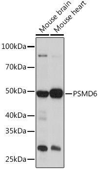
Western blot analysis of various lysates using PSMD6 Rabbit pAb (CAB18263) at 1:1000 dilution. Secondary antibody: HRP-conjugated Goat anti-Rabbit IgG (H+L) (CABS014) at 1:10000 dilution. Lysates/proteins: 25μg per lane. Blocking buffer: 3% nonfat dry milk in TBST. Detection: ECL Basic Kit (AbGn00020). Exposure time: 3min.
Western blot analysis of lysates from Rat brain, using PSMD6 Rabbit pAb (CAB18263) at 1:1000 dilution. Secondary antibody: HRP-conjugated Goat anti-Rabbit IgG (H+L) (CABS014) at 1:10000 dilution. Lysates/proteins: 25μg per lane. Blocking buffer: 3% nonfat dry milk in TBST. Detection: ECL Enhanced Kit (AbGn00021). Exposure time: 90s.