The REG3G Antibody (CAB2146) is a high-quality antibody developed for reliable detection and analysis of target proteins. This antibody, generated in rabbits, is highly specific to human samples and is validated for use in various applications including Western blotting.REG3G is a member of the regenerating islet-derived (REG) gene family and is involved in the regulation of immune responses and inflammation.
This antibody is validated for use in WB, IF/ICC, ELISA applications and has demonstrated reactivity against Human, Mouse, Rat samples.
Product Name:
REG3G Antibody
SKU:
CAB2146
Size:
20μL, 100μL
Reactivity:
Human, Mouse, Rat
Conjugate:
Unconjugated
Immunogen:
Synthetic peptide. This information is considered to be commercially sensitive.
Recommended starting concentration is 1 μg/mL. Please optimize the concentration based on your specific assay requirements.
Synonyms:
REG-3-gamma, reg III-gamma, REG3G
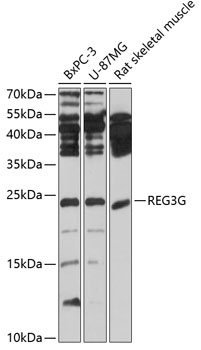
Positive Sample:
BxPC-3, U-87MG, Rat skeletal muscle
Cellular Localization:
Cytoplasm, Secreted.
Calculated MW:
19kDa
Observed MW:
19kDa
This gene encodes a C-type lectin that demonstrates bactericidal activity. This gene is predominantly expressed in the distal small intestine where the encoded protein undergoes proteolytic processing by trypsin. Mice lacking the encoded protein exhibit altered mucus distribution, increased bacterial contact with the epithelium, and elevated inflammatory markers in the ileum, and low-grade inflammation.
Purification Method
Affinity purification
Gene ID
19695
RRID
AB_2764165
Buffer Information
Store at -20℃. Avoid freeze / thaw cycles. Buffer: PBS containing 50% glycerol, preserved with proclin300 or sodium azide, pH 7.3.
Western blot analysis of various lysates using REG3G Rabbit pAb (CAB2146) at 1:1000 dilution. Secondary antibody: HRP-conjugated Goat anti-Rabbit IgG (H+L) (CABS014) at 1:10000 dilution. Lysates/proteins: 25μg per lane. Blocking buffer: 3% nonfat dry milk in TBST. Detection: ECL Basic Kit (AbGn00020). Exposure time: 10s.
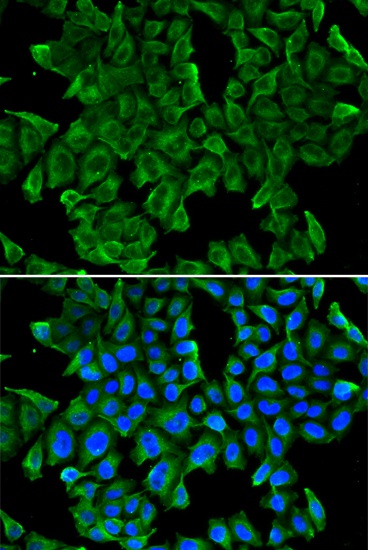
Immunofluorescence analysis of MCF-7 cells using REG3G Rabbit pAb (CAB2146). Secondary antibody: Cy3-conjugated Goat anti-Rabbit IgG (H+L) (CABS007) at 1:500 dilution. Blue: DAPI for nuclear staining.