The RPL17 Antibody (CAB5934) is a high-quality antibody developed for reliable detection and analysis of target proteins. This antibody, generated in rabbits, is highly specific for human samples and has been validated for use in Western blot applications. By binding to the RPL17 protein, the antibody allows for the detection and analysis of RPL17 in a variety of cell types, making it ideal for studies in molecular biology and cancer research.RPL17 is a crucial component of the ribosome, the cellular machinery responsible for protein production.
This antibody is validated for use in WB, IHC-P, IF/ICC, ELISA applications and has demonstrated reactivity against Human, Mouse, Rat samples.
Product Name:
RPL17 Antibody
SKU:
CAB5934
Size:
20μL, 100μL
Reactivity:
Human, Mouse, Rat
Conjugate:
Unconjugated
Immunogen:
Recombinant protein (or fragment).This information is considered to be commercially sensitive.
Ribosomes, the organelles that catalyze protein synthesis, consist of a small 40S subunit and a large 60S subunit. Together these subunits are composed of 4 RNA species and approximately 80 structurally distinct proteins. This gene encodes a ribosomal protein that is a component of the 60S subunit. The protein belongs to the L22P family of ribosomal proteins. It is located in the cytoplasm. This gene has been referred to as rpL23 because the encoded protein shares amino acid identity with ribosomal protein L23 from Halobacterium marismortui; however, its official symbol is RPL17. As is typical for genes encoding ribosomal proteins, there are multiple processed pseudogenes of this gene dispersed through the genome. Alternative splicing results in multiple transcript variants. Read-through transcription also exists between this gene and the neighboring downstream C18orf32 (chromosome 18 open reading frame 32) gene.
Purification Method
Affinity purification
Gene ID
6139
RRID
AB_2766671
Buffer Information
Store at -20℃. Avoid freeze / thaw cycles. Buffer: PBS containing 50% glycerol, preserved with proclin300 or sodium azide, pH 7.3.
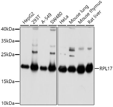
Western blot analysis of various lysates using RPL17 Rabbit pAb (CAB5934) at 1:1000 dilution. Secondary antibody: HRP-conjugated Goat anti-Rabbit IgG (H+L) (CABS014) at 1:10000 dilution. Lysates/proteins: 25μg per lane. Blocking buffer: 3% nonfat dry milk in TBST. Detection: ECL Basic Kit (AbGn00020). Exposure time: 5s.
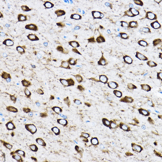
Immunohistochemistry analysis of paraffin-embedded Rat brain using RPL17 Rabbit pAb (CAB5934) at dilution of 1:100 (40x lens). High pressure antigen retrieval performed with 0.01M Citrate buffer (pH 6.0) prior to IHC staining.
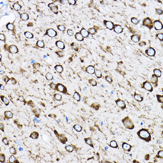
Immunohistochemistry analysis of paraffin-embedded Mouse brain using RPL17 Rabbit pAb (CAB5934) at dilution of 1:100 (40x lens). High pressure antigen retrieval performed with 0.01M Citrate buffer (pH 6.0) prior to IHC staining.
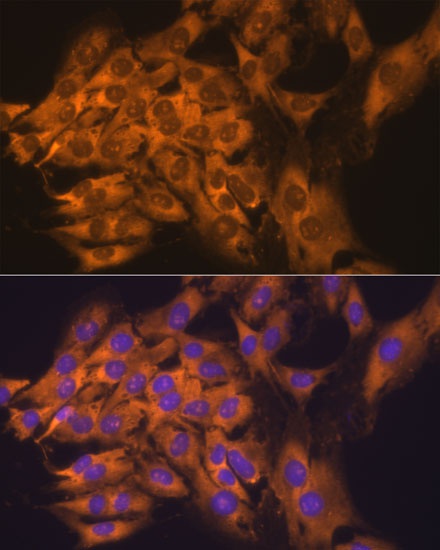
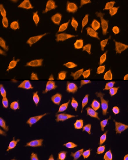
Immunofluorescence analysis of C6 cells using RPL17 Rabbit pAb (CAB5934) at dilution of 1:100. Secondary antibody: Cy3-conjugated Goat anti-Rabbit IgG (H+L) (CABS007) at 1:500 dilution. Blue: DAPI for nuclear staining.
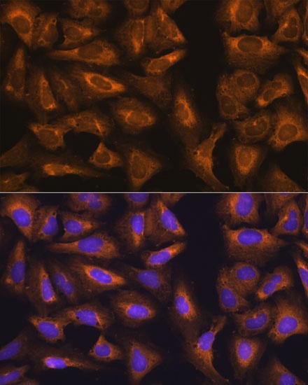
Immunofluorescence analysis of L929 cells using RPL17 Rabbit pAb (CAB5934) at dilution of 1:100. Secondary antibody: Cy3-conjugated Goat anti-Rabbit IgG (H+L) (CABS007) at 1:500 dilution. Blue: DAPI for nuclear staining.
Immunofluorescence analysis of U-2 OS cells using RPL17 Rabbit pAb (CAB5934) at dilution of 1:100. Secondary antibody: Cy3-conjugated Goat anti-Rabbit IgG (H+L) (CABS007) at 1:500 dilution. Blue: DAPI for nuclear staining.