The Sheep Estrogen ELISA Kit is a reliable and accurate tool for detecting estrogen levels in sheep serum, plasma, and cell culture supernatants. With high sensitivity and specificity, this kit provides precise and reproducible results, making it ideal for various research applications. Estrogen is a vital hormone in sheep physiology, playing a crucial role in reproductive function and overall health.
Monitoring estrogen levels is important for understanding sheep reproduction, managing breeding programs, and studying reproductive diseases. This ELISA kit is easy to use and offers a fast and efficient way to measure estrogen levels in sheep samples. Researchers and veterinarians can rely on this kit to support their studies on sheep reproduction, hormone regulation, and fertility management.
Product Name:
Sheep Estrogen ELISA Kit
SKU:
SHFI00063
Reactivity:
Sheep
Assay Type:
Competitive ELISA, Coated with Antigen
Sensitivity:
9.375 pg/mL
Range:
15.625-1000 pg/mL
Sample Type:
Serum, Plasma, Cell Culture Supernatant, Cell or Tissue Lysate, Other Liquid Samples
Storage:
2-8°C(Sealed), Don't cryopreserve.
Linearity:
Sample
1:2
1:4
1:8
Serum (n = 10)
87-104%
86-100%
90-96%
EDTA Plasma (n = 10)
88-97%
83-99%
85-101%
Heparin Plasma (n = 10)
82-92%
84-100%
84-100%
Recovery:
Sample
Recovery Range (%)
Average (%)
Serum (n = 10)
87-100
92
EDTA Plasma (n = 10)
85-105
96
Heparin Plasma (n = 10)
85-105
96
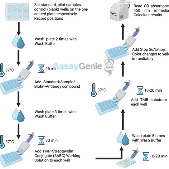
Note: The below protocol is a sample protocol. Protocols are specific to each batch/lot. For the correct instructions please follow the protocol included in your kit.
Step
Procedure
1
Reagent & Plate Preparation: Equilibrate TMB substrate for 30 minutes at room temperature. Prepare standards, samples (minimum 1:2 dilution), blanks, assign wells, and pre-wash the plate twice.
2
Sample & Biotin-Antibody Binding: Add 50 µL standard or sample followed by 50 µL biotin-labeled antibody to each well. Mix gently and incubate at 37°C for 45 minutes.
3
Washing: Wash the plate 5 times with wash buffer, allowing 1 minute soak time per wash.
4
Color Development: Add TMB substrate and incubate in the dark at 37°C for 10-20 minutes until color develops.
5
Stop Reaction: Add stop solution to terminate the reaction. The color changes from blue to yellow immediately.
6
Reading: Measure absorbance at 450 nm using a microplate reader.
Sample Type
Protocol
Serum
Allow blood to clot, centrifuge at 1000 × g for 20 minutes, collect supernatant and store appropriately.
Plasma
Collect using EDTA anticoagulant, centrifuge at 1000 × g for 15 minutes at 2–8°C and collect plasma.
Cell Culture Supernatant
Centrifuge at 1000 × g for 20 minutes at 4°C and collect clarified supernatant.
Cell Lysate
Lyse cells using recommended lysis buffer with protease inhibitors, centrifuge at 10,000 rpm for 10 minutes, and collect protein supernatant.
Tissue Homogenate
Homogenize tissue in PBS with protease inhibitors, centrifuge at 5000 × g for 5 minutes, and collect supernatant.
Other Sample Types
Centrifuge samples at 1000 × g for 15 minutes at 2–8°C and collect supernatant. For additional guidance, please contact techsupport@assaygenie.com.
Component
Quantity
Storage
48T
96T
ELISA Microplate (Dismountable)
8×6
8×12
Place the test strips into a sealed foil bag with the desiccant. Store for 1 month at 2-8°C; Store for 12 months at -20°C.
Lyophilized Standard
1 vial
2 vial
Place the standards into a sealed foil bag with the desiccant. Store for 1 month at 2-8°C; Store for 12 months at -20°C.
Biotin-labeled Antibody (Lyophilized)
1 vial
1 vial
Place the standards into a sealed foil bag with the desiccant. Store for 1 month at 2-8°C; Store for 12 months at -20°C.
HRP-Streptavidin Conjugate (SABC, 100X)
60 ul
120 ul
2-8°C (Avoid direct light)
TMB Substrate
5 ml
10 ml
2-8°C (Avoid direct light)
Purified Water
200 ul
200 ul
2-8°C
Sample Dilution Buffer
10 ml
20 ml
2-8°C
Antibody Dilution Buffer
5 ml
10 ml
2-8°C
SABC Dilution Buffer
5 ml
10 ml
2-8°C
Stop Solution
5 ml
5 ml
2-8°C
Wash Buffer(25X)
15 ml
30 ml
2-8°C
Plate Sealer
3 pieces
5 pieces
-
Technical Manual
1 copy
1 copy
-
Tsiartas et al.
Seven days ex vivo perfusion of whole ewe ovaries with follicular maturation and oocyte retrieval: towards the development of an alternative fertility preservation method


")
")

")

")