The SOD2 Polyclonal Antibody (CAB21805) is a high-quality antibody developed for reliable detection and analysis of target proteins. This antibody, produced in rabbits, has a high reactivity with human samples and is validated for use in Western blot applications. By binding to the SOD2 protein, this antibody allows for the detection and analysis of SOD2 in a variety of cell types, making it an essential component in studies related to oxidative stress, cancer, and other diseases.SOD2, also known as manganese superoxide dismutase, is an essential enzyme that helps to protect cells from damage caused by reactive oxygen species.
This antibody is validated for use in WB, IHC-P, IF/ICC, ELISA applications and has demonstrated reactivity against Human, Mouse, Rat samples.
Product Name:
SOD2 Polyclonal Antibody
SKU:
CAB21805
Size:
20μL, 100μL
Reactivity:
Human, Mouse, Rat
Conjugate:
Unconjugated
Immunogen:
Recombinant protein (or fragment).This information is considered to be commercially sensitive.
HeLa, 293T, Hep G2, NIH/3T3, RAW 264.7, 293T, Mouse heart, Rat brain, Rat heart
Cellular Localization:
Mitochondrion Matrix.
Calculated MW:
25kDa
Observed MW:
Refertofigures
This gene is a member of the iron/manganese superoxide dismutase family. It encodes a mitochondrial protein that forms a homotetramer and binds one manganese ion per subunit. This protein binds to the superoxide byproducts of oxidative phosphorylation and converts them to hydrogen peroxide and diatomic oxygen. Mutations in this gene have been associated with idiopathic cardiomyopathy (IDC), premature aging, sporadic motor neuron disease, and cancer. Alternative splicing of this gene results in multiple transcript variants. A related pseudogene has been identified on chromosome 1.
Purification Method
Affinity purification
Gene ID
6648
Buffer Information
Store at -20℃. Avoid freeze / thaw cycles. Buffer: PBS containing 50% glycerol, preserved with proclin300 or sodium azide, pH 7.3.
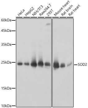
Western blot analysis of various lysates, using SOD2 Rabbit pAb (CAB21805) at 1:1000 dilution. Secondary antibody: HRP-conjugated Goat anti-Rabbit IgG (H+L) (CABS014) at 1:10000 dilution. Lysates/proteins: 25μg per lane. Blocking buffer: 3% nonfat dry milk in TBST. Detection: ECL Basic Kit (AbGn00020). Exposure time: 1s.
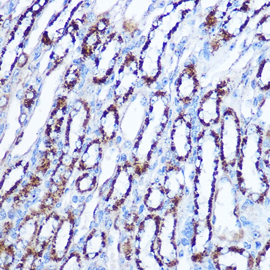
Immunohistochemistry analysis of paraffin-embedded Rat kidney using SOD2 Rabbit pAb (CAB21805) at dilution of 1:100 (40x lens). Microwave antigen retrieval performed with 0.01M Tris/EDTA Buffer (pH 9.0) prior to IHC staining.
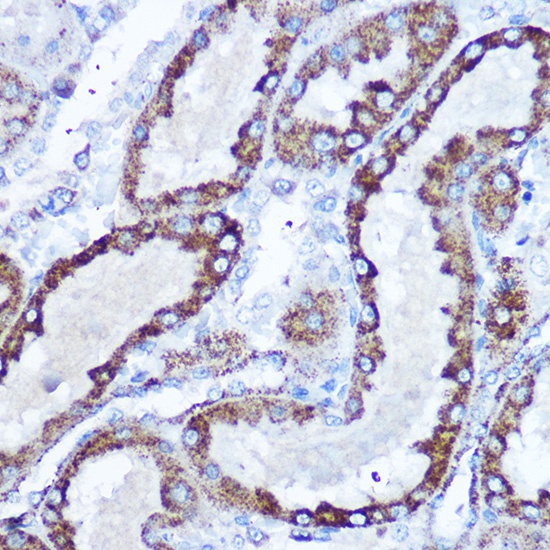
Immunohistochemistry analysis of paraffin-embedded Mouse kidney using SOD2 Rabbit pAb (CAB21805) at dilution of 1:100 (40x lens). Microwave antigen retrieval performed with 0.01M Tris/EDTA Buffer (pH 9.0) prior to IHC staining.
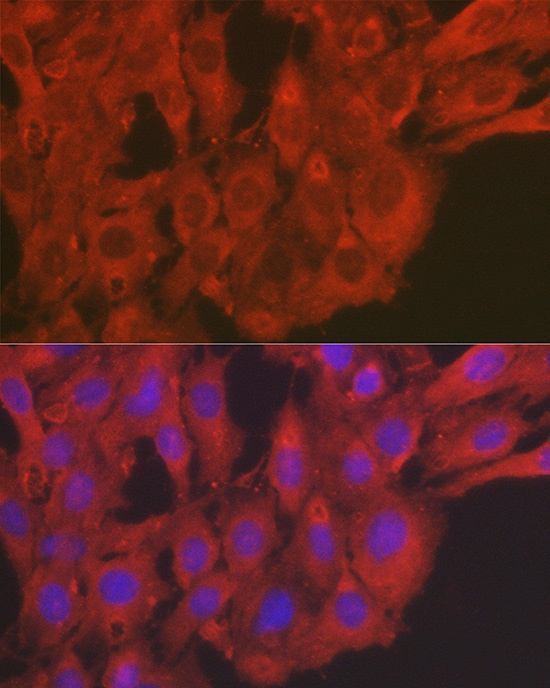
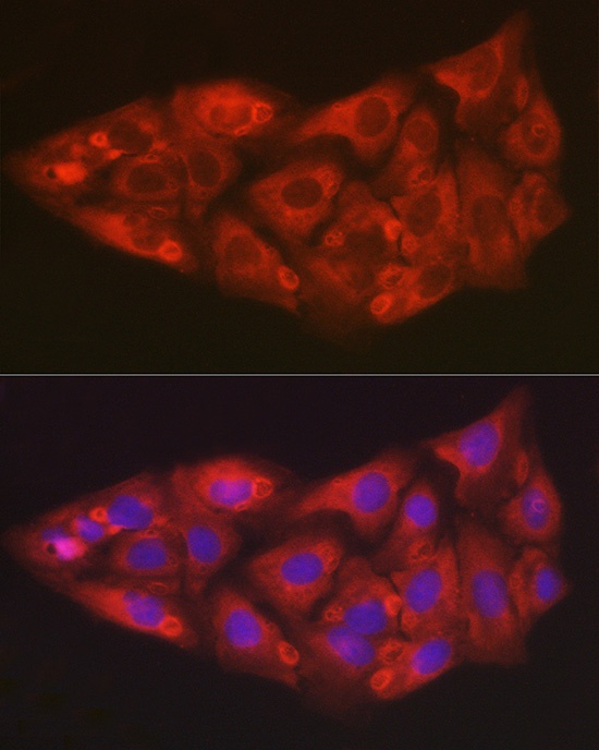
Immunofluorescence analysis of C6 cells using SOD2 Rabbit pAb (CAB21805) at dilution of 1:50 (40x lens). Secondary antibody: Cy3-conjugated Goat anti-Rabbit IgG (H+L) (CABS007) at 1:500 dilution. Blue: DAPI for nuclear staining.
Immunofluorescence analysis of U2OS cells using SOD2 Rabbit pAb (CAB21805) at dilution of 1:50 (40x lens). Secondary antibody: Cy3-conjugated Goat anti-Rabbit IgG (H+L) (CABS007) at 1:500 dilution. Blue: DAPI for nuclear staining.
 at 1:1000 dilution. Secondary antibody: HRP Goat Anti-Rabbit IgG (H+L) at 1:10000 dilution. Lysates/proteins: 25ug per lane. Blocking buffer: 3% nonfat dry milk in TBST.")
 at 1:1000 dilution. Secondary antibody: HRP Goat Anti-Rabbit IgG (H+L) at 1:10000 dilution. Lysates/proteins: 25ug per lane. Blocking buffer: 3% nonfat dry milk in TBST.")

![Anti-SOD2 [R67-7H-3] Monoclonal Antibody (AGMB01762)](https://cdn11.bigcommerce.com/s-h68l9z2lnx/images/stencil/590x590/products/273051/677512/anti-sod2-r67-7h-3-monoclonal-antibody-agmb01762__62651.1773032801.jpg?c=2 "Anti-SOD2 [R67-7H-3] Monoclonal Antibody (AGMB01762)")
![Anti- SOD2 [1H6] Monoclonal Antibody - Knockout Validated (AGMB06742)](https://cdn11.bigcommerce.com/s-h68l9z2lnx/images/stencil/590x590/products/278023/731356/anti-sod2-1h6-monoclonal-antibody-knockout-validated-agmb06742__19594.1777183413.jpg?c=2 "Anti- SOD2 [1H6] Monoclonal Antibody - Knockout Validated (AGMB06742)")

![Anti-SOD2 (4C4) [4C4-G8-H12] Monoclonal Antibody (AGMB04366)](https://cdn11.bigcommerce.com/s-h68l9z2lnx/images/stencil/590x590/products/275655/677086/anti-sod2-4c4-4c4-g8-h12-monoclonal-antibody-agmb04366__29039.1773031469.jpg?c=2 "Anti-SOD2 (4C4) [4C4-G8-H12] Monoclonal Antibody (AGMB04366)")