The TAT Antibody (CAB6764) is a high-quality antibody developed for reliable detection and analysis of target proteins. This antibody is produced in rabbits and has been rigorously tested for its reactivity with human samples, making it an excellent choice for Western blot applications.The TAT protein is involved in a variety of cellular processes, including gene expression and cell differentiation, making it a crucial target for research in fields such as molecular biology and cancer biology. By specifically binding to the TAT protein, this antibody enables precise detection and analysis of TAT in various cell types, providing valuable insights into its functions and mechanisms of action.
This antibody is validated for use in WB, IHC-P, ELISA applications and has demonstrated reactivity against Human, Mouse, Rat samples.
Product Name:
TAT Antibody
SKU:
CAB6764
Size:
20μL, 100μL
Reactivity:
Human, Mouse, Rat
Conjugate:
Unconjugated
Immunogen:
Recombinant protein (or fragment).This information is considered to be commercially sensitive.
This nuclear gene encodes a mitochondrial protein tyrosine aminotransferase which is present in the liver and catalyzes the conversion of L-tyrosine into p-hydroxyphenylpyruvate. Mutations in this gene cause tyrosinemia (type II, Richner-Hanhart syndrome), a disorder accompanied by major skin and corneal lesions, with possible cognitive disability. A regulator gene for tyrosine aminotransferase is X-linked.
Purification Method
Affinity purification
Gene ID
6898
RRID
AB_2767347
Buffer Information
Store at -20℃. Avoid freeze / thaw cycles. Buffer: PBS containing 50% glycerol, preserved with proclin300 or sodium azide, pH 7.3.
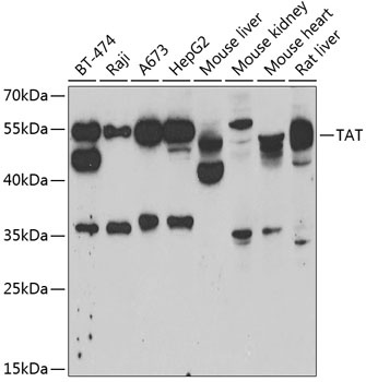
Western blot analysis of various lysates using TAT Rabbit pAb (CAB6764) at 1:1000 dilution. Secondary antibody: HRP-conjugated Goat anti-Rabbit IgG (H+L) (CABS014) at 1:10000 dilution. Lysates/proteins: 25μg per lane. Blocking buffer: 3% nonfat dry milk in TBST. Detection: ECL Basic Kit (AbGn00020). Exposure time: 5min.
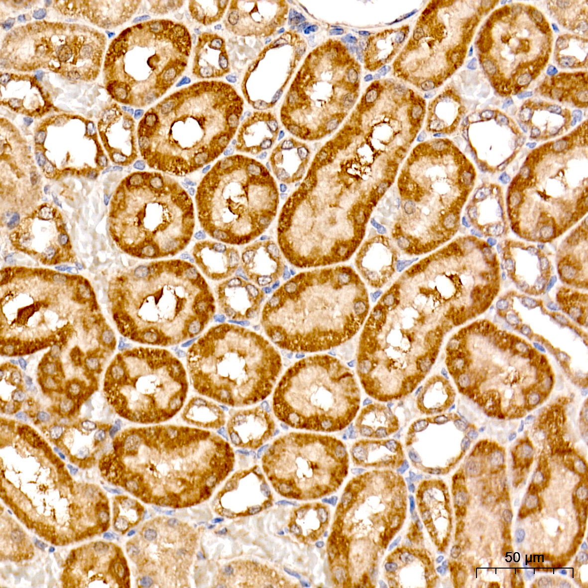
Immunohistochemistry analysis of paraffin-embedded Rat kidney tissue using TAT Rabbit pAb (CAB6764) at a dilution of 1:500 (40x lens). High pressure antigen retrieval was performed with 0.01 M citrate buffer (pH 6.0) prior to IHC staining.