The THRSP Antibody (CAB7232) is a high-quality antibody developed for reliable detection and analysis of target proteins. This antibody, produced in rabbits, exhibits high reactivity with human samples and is specifically validated for use in Western blot applications. It binds to the THRSP protein, allowing for accurate detection and analysis in different cell types.THRSP is an important regulator of lipid metabolism and is involved in the synthesis and storage of fatty acids in the body. Its role in lipid homeostasis makes it a key target for studies related to metabolic disorders, obesity, and diabetes.
This antibody is validated for use in WB, IF/ICC, ELISA applications and has demonstrated reactivity against Human, Mouse, Rat samples.
Product Name:
THRSP Antibody
SKU:
CAB7232
Size:
20μL, 100μL
Reactivity:
Human, Mouse, Rat
Conjugate:
Unconjugated
Immunogen:
Recombinant protein (or fragment).This information is considered to be commercially sensitive.
Recommended starting concentration is 1 μg/mL. Please optimize the concentration based on your specific assay requirements.
Synonyms:
S14, Lpgp, THRP, LPGP1, SPOT14, THRSP
Positive Sample:
293T
Cellular Localization:
Cytoplasm, Nucleus.
Calculated MW:
17kDa
Observed MW:
20kDa
The protein encoded by this gene is similar to the gene product of S14, a rat gene whose expression is limited to liver and adipose tissue and is controlled by nutritional and hormonal factors. This gene has been shown to be expressed in liver and adipocytes, particularly in lipomatous modules. It is also found to be expressed in lipogenic breast cancers, which suggests a role in controlling tumor lipid metabolism.
Purification Method
Affinity purification
Gene ID
7069
RRID
AB_2767780
Buffer Information
Store at -20℃. Avoid freeze / thaw cycles. Buffer: PBS containing 50% glycerol, preserved with proclin300 or sodium azide, pH 7.3.
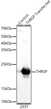
Western blot analysis of lysates from wild type (WT) and 293T cells transfected with THRSP using THRSP Rabbit pAb (CAB7232) at 1:500 dilution. Secondary antibody: HRP-conjugated Goat anti-Rabbit IgG (H+L) (CABS014) at 1:10000 dilution. Lysates/proteins: 25μg per lane. Blocking buffer: 3% nonfat dry milk in TBST. Detection: ECL Basic Kit (AbGn00020). Exposure time: 10s.
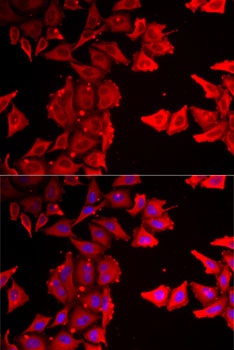
Immunofluorescence analysis of MCF7 cells using THRSP Rabbit pAb (CAB7232). Secondary antibody: Cy3-conjugated Goat anti-Rabbit IgG (H+L) (CABS007) at 1:500 dilution. Blue: DAPI for nuclear staining.