The TMF1 Antibody (CAB17542) is a high-quality antibody developed for reliable detection and analysis of target proteins. This rabbit polyclonal antibody is highly specific for human samples and is validated for use in Western blot applications. By binding to the TMF1 protein, this antibody enables detection and analysis in a variety of cell types, making it an essential tool for studies in cell biology and molecular research.TMF1, also known as TATA element modulatory factor 1, is involved in diverse cellular processes such as transcriptional regulation, protein degradation, and cellular stress responses.
This antibody is validated for use in WB, ELISA applications and has demonstrated reactivity against Human, Mouse samples.
Product Name:
TMF1 Antibody
SKU:
CAB17542
Size:
20μL, 100μL
Reactivity:
Human, Mouse
Conjugate:
Unconjugated
Immunogen:
Synthetic peptide. This information is considered to be commercially sensitive.
Recommended starting concentration is 1 μg/mL. Please optimize the concentration based on your specific assay requirements.
Synonyms:
TMF, ARA160, TMF1
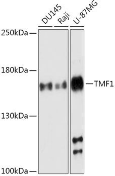
Positive Sample:
DU145, Raji, U-87MG
Cellular Localization:
Cytoplasm, Golgi Apparatus Membrane, Nucleus.
Calculated MW:
123kDa
Observed MW:
160kDa
Enables androgen receptor binding activity and nuclear receptor coactivator activity. Involved in androgen receptor signaling pathway and positive regulation of transcription by RNA polymerase II. Located in Golgi apparatus.
Purification Method
Affinity purification
Gene ID
7110
RRID
AB_2772644
Buffer Information
Store at -20℃. Avoid freeze / thaw cycles. Buffer: PBS with 0.01% thimerosal,50% glycerol,pH7.3.
Western blot analysis of various lysates using TMF1 Rabbit pAb (CAB17542) at 1:1000 dilution. Secondary antibody: HRP-conjugated Goat anti-Rabbit IgG (H+L) (CABS014) at 1:10000 dilution. Lysates/proteins: 25μg per lane. Blocking buffer: 3% nonfat dry milk in TBST. Detection: ECL Basic Kit (AbGn00020). Exposure time: 180s.