 at1:1000 dilution. THP-1 cells were treated by LPS (1 μg/ml) at 37℃ for 8 hours. Secondary antibody: HRP Goat Anti-Rabbit IgG (H+L) at 1:10000 dilution. Lysates/proteins: 25μg per lane. Blocking buffer: 3% nonfat dry milk in TBST.")
Description
TNF-alpha Monoclonal Antibody (CAB22227)
The TNF-alpha Monoclonal Antibody (CAB22227) is a high-quality antibody developed for reliable detection and analysis of target proteins. This antibody, produced through advanced monoclonal technology, recognizes and binds specifically to TNF in human samples, making it an essential reagent for various immunological and cancer-related studies.TNF is a critical regulator of inflammatory processes and has been implicated in a wide range of diseases, including autoimmune disorders, infectious diseases, and cancer. By targeting TNF with this monoclonal antibody, researchers can gain important insights into the role of TNF in disease pathogenesis and potentially identify new therapeutic strategies for TNF-associated conditions.
This antibody is validated for use in WB, IF/ICC, IP, ELISA applications and has demonstrated reactivity against Human samples.
| Product Name: | TNF-alpha Monoclonal Antibody |
| SKU: | CAB22227 |
| Size: | 20μL, 100μL |
| Reactivity: | Human |
| Clone Number: | ARC52531 |
| Conjugate: | Unconjugated |
| Immunogen: | Recombinant protein (or fragment).This information is considered to be commercially sensitive. | ||||||||
| Sequence: | VRSS SRTP SDKP VAHV VANP QAEG QLQW LNRR ANAL LANG VELR DNQL VVPS EGLY LIYS QVLF KGQG CPST HVLL THTI SRIA VSYQ TKVN LLSA IKSP CQRE TPEG AEAK PWYE PIYL GGVF QLEK GDRL SAEI NRPD YLDF AESG QVYF GIIA L | ||||||||
| Tested Applications: | WB IF/ICC IP ELISA | ||||||||
| Recommended Dilution: |
| ||||||||
| Synonyms: | DIF, TNFA, TNFSF2, TNLG1F, TNF-alpha, TNF-α |
| Positive Sample: | THP-1 treated with LPS; THP-1 treated with IFN-γ, LPS and BFA |
| Cellular Localization: | Cell Membrane, Membrane, Secreted. |
| Calculated MW: | 26kDa |
| Observed MW: | 18kDa/25kDa |
This gene encodes a multifunctional proinflammatory cytokine that belongs to the tumor necrosis factor (TNF) superfamily. This cytokine is mainly secreted by macrophages. It can bind to, and thus functions through its receptors TNFRSF1A/TNFR1 and TNFRSF1B/TNFBR. This cytokine is involved in the regulation of a wide spectrum of biological processes including cell proliferation, differentiation, apoptosis, lipid metabolism, and coagulation. This cytokine has been implicated in a variety of diseases, including autoimmune diseases, insulin resistance, psoriasis, rheumatoid arthritis ankylosing spondylitis, tuberculosis, autosomal dominant polycystic kidney disease, and cancer. Mutations in this gene affect susceptibility to cerebral malaria, septic shock, and Alzheimer disease. Knockout studies in mice also suggested the neuroprotective function of this cytokine.
| Purification Method | Affinity purification |
| Gene ID | 7124 |
| Buffer Information | Store at -20℃. Avoid freeze / thaw cycles. Buffer: PBS with 0.09% sodium azide,0.05% BSA,50% glycerol,pH7.3. |
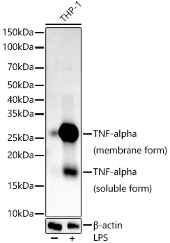 | Western blot analysis of various lysates, using TNF-α Rabbit mAb (CAB22227) at1:34000 dilution. THP-1 cells were treated with LPS (1 μg/ml) at 37℃ for 8 hours. Secondary antibody: HRP-conjugated Goat anti-Rabbit IgG (H+L) (CABS014) at 1:10000 dilution. Lysates/proteins: 25μg per lane. Blocking buffer: 3% nonfat dry milk in TBST. Detection: ECL Basic Kit (AbGn00020). Exposure time: 90s. |
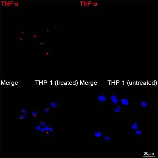 | Confocal imaging of THP-1 cells (treated with TPA and LPS) and THP-1 cells (untreated) using TNF-α Rabbit mAb (CAB22227, dilution 1:3000) followed by a further incubation with Cy3-conjugated Goat Anti-Rabbit IgG (H+L) (CABS007, dilution 1:500) (Red). DAPI was used for nuclear staining (Blue). Objective: 100x. |
 at1:1000 dilution. THP-1 cells were treated by LPS (1 μg/ml) at 37℃ for 8 hours. Secondary antibody: HRP Goat Anti-Rabbit IgG (H+L) at 1:10000 dilution. Lysates/proteins: 25μg per lane. Blocking buffer: 3% nonfat dry milk in TBST.")

![Anti-TNF alpha [7A8] Monoclonal Antibody, capture (AGMB05977)](https://cdn11.bigcommerce.com/s-h68l9z2lnx/images/stencil/590x590/products/277261/677360/anti-tnf-alpha-7a8-monoclonal-antibody-capture-agmb05977__06328.1773032328.jpg?c=2 "Anti-TNF alpha [7A8] Monoclonal Antibody, capture (AGMB05977)")
![Anti-TNF alpha [5F11] Monoclonal Antibody, unconjugated, detector (AGMB05978)](https://cdn11.bigcommerce.com/s-h68l9z2lnx/images/stencil/590x590/products/277262/679393/anti-tnf-alpha-5f11-monoclonal-antibody-unconjugated-detector-agmb05978__53771.1773038783.jpg?c=2 "Anti-TNF alpha [5F11] Monoclonal Antibody, unconjugated, detector (AGMB05978)")
![Anti-TNF alpha (9D9) [9D9-6A9-8B4] Monoclonal Antibody (AGMB04386)](https://cdn11.bigcommerce.com/s-h68l9z2lnx/images/stencil/590x590/products/275675/676167/anti-tnf-alpha-9d9-9d9-6a9-8b4-monoclonal-antibody-agmb04386__89518.1773028586.jpg?c=2 "Anti-TNF alpha (9D9) [9D9-6A9-8B4] Monoclonal Antibody (AGMB04386)")
![Anti-TNF alpha (6H2) [6H2-4C6-5B6] Monoclonal Antibody (AGMB04385)](https://cdn11.bigcommerce.com/s-h68l9z2lnx/images/stencil/590x590/products/275674/678080/anti-tnf-alpha-6h2-6h2-4c6-5b6-monoclonal-antibody-agmb04385__71977.1773034596.jpg?c=2 "Anti-TNF alpha (6H2) [6H2-4C6-5B6] Monoclonal Antibody (AGMB04385)")