The TPST2 Polyclonal Antibody (CAB18141) is a high-quality antibody developed for reliable detection and analysis of target proteins. This rabbit-raised antibody is highly specific to TPST2 in human samples and is suitable for use in Western blot applications. By targeting the TPST2 protein, this antibody allows for precise detection and analysis in various cell types, making it ideal for investigations in areas such as glycobiology and protein modification.TPST2 is known for its role in post-translational modification of proteins through tyrosine sulfation, which is crucial for their proper function in processes like cell adhesion and signaling.
This antibody is validated for use in WB, ELISA applications and has demonstrated reactivity against Human, Mouse samples.
Product Name:
TPST2 Polyclonal Antibody
SKU:
CAB18141
Size:
20μL, 100μL
Reactivity:
Human, Mouse
Conjugate:
Unconjugated
Immunogen:
Recombinant protein (or fragment).This information is considered to be commercially sensitive.
Recommended starting concentration is 1 μg/mL. Please optimize the concentration based on your specific assay requirements.
Synonyms:
TPST-2, TANGO13B, TPST2
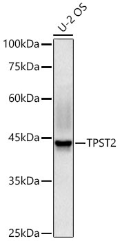
Positive Sample:
U-2 OS
Cellular Localization:
Golgi Apparatus Membrane, Single-Pass Type Ii Membrane Protein.
Calculated MW:
42kDa
Observed MW:
42kDa
The protein encoded by this gene catalyzes the O-sulfation of tyrosine residues within acidic regions of proteins. The encoded protein is a type II integral membrane protein found in the Golgi body. Alternative splicing produces multiple transcript variants encoding distinct isoforms.
Purification Method
Affinity purification
Gene ID
8459
RRID
AB_2861932
Buffer Information
Store at -20℃. Avoid freeze / thaw cycles. Buffer: PBS containing 50% glycerol, preserved with proclin300 or sodium azide, pH 7.3.
Western blot analysis of lysates from U-2 OS cells using TPST2 Rabbit pAb (CAB18141) at 1:1000 dilution incubated overnight at 4°C. Secondary antibody: HRP-conjugated Goat anti-Rabbit IgG (H+L) (CABS014) at 1:10000 dilution. Lysates/proteins: 25 μg per lane. Blocking buffer: 3% nonfat dry milk in TBST. Detection: ECL Basic Kit (AbGn00020) Exposure time: 60s.