The VAPA Antibody (CAB12939) is a high-quality antibody developed for reliable detection and analysis of target proteins. This antibody, produced in rabbits, exhibits strong reactivity with human samples and is validated for use in Western blot assays. By specifically binding to VAPA, researchers can effectively detect and analyze this protein in a variety of cell types, making it well-suited for investigations in cell biology and neurobiology.
This antibody is validated for use in WB, IHC-P, ELISA applications and has demonstrated reactivity against Human, Mouse samples.
Product Name:
VAPA Antibody
SKU:
CAB12939
Size:
20μL, 100μL
Reactivity:
Human, Mouse
Conjugate:
Unconjugated
Immunogen:
Recombinant protein (or fragment).This information is considered to be commercially sensitive.
Recommended starting concentration is 1 μg/mL. Please optimize the concentration based on your specific assay requirements.
Synonyms:
VAP-A, VAP33, VAMP-A, VAP-33, hVAP-33, VAPA
Positive Sample:
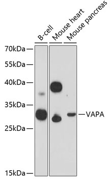
B-cell, Mouse heart, Mouse pancreas
Cellular Localization:
Endoplasmic Reticulum Membrane, Single-Pass Type Iv Membrane Protein.
Calculated MW:
28kDa
Observed MW:
28kDa
The protein encoded by this gene is a type IV membrane protein. It is present in the plasma membrane and intracellular vesicles. It may also be associated with the cytoskeleton. This protein may function in vesicle trafficking, membrane fusion, protein complex assembly and cell motility. Alternative splicing occurs at this locus and two transcript variants encoding distinct isoforms have been identified.
Purification Method
Affinity purification
Gene ID
9218
RRID
AB_2759785
Buffer Information
Store at -20℃. Avoid freeze / thaw cycles. Buffer: PBS with 0.01% thimerosal,50% glycerol,pH7.3.
Western blot analysis of various lysates using VAPA Rabbit pAb (CAB12939) at 1:3000 dilution. Secondary antibody: HRP-conjugated Goat anti-Rabbit IgG (H+L) (CABS014) at 1:10000 dilution. Lysates/proteins: 25μg per lane. Blocking buffer: 3% nonfat dry milk in TBST. Detection: ECL Enhanced Kit (AbGn00021). Exposure time: 90s.
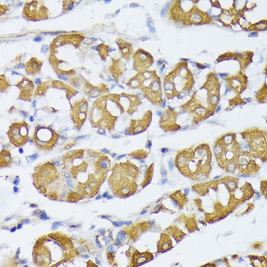
Immunohistochemistry analysis of paraffin-embedded Human stomach using VAPA Rabbit pAb (CAB12939) at dilution of 1:100 (40x lens). Microwave antigen retrieval performed with 0.01M PBS Buffer (pH 7.2) prior to IHC staining.