The WNK3 Antibody (CAB15507) is a high-quality antibody developed for reliable detection and analysis of target proteins. This antibody, produced in rabbits, exhibits high reactivity with human samples and has been validated for use in Western blot applications. By specifically binding to the WNK3 protein, this antibody allows for precise detection and analysis in various cell types, making it an essential component for studies in physiology and cardiovascular research.
This antibody is validated for use in WB, IF/ICC, ELISA applications and has demonstrated reactivity against Human, Mouse, Rat samples.
Product Name:
WNK3 Antibody
SKU:
CAB15507
Size:
20μL, 100μL
Reactivity:
Human, Mouse, Rat
Conjugate:
Unconjugated
Immunogen:
Recombinant protein (or fragment).This information is considered to be commercially sensitive.
Recommended starting concentration is 1 μg/mL. Please optimize the concentration based on your specific assay requirements.
Synonyms:
PRKWNK3, WNK3
Positive Sample:
HAP1
Cellular Localization:
Cytoplasm.
Calculated MW:
198kDa
Observed MW:
180kDa
This gene encodes a protein belonging to the 'with no lysine' family of serine-threonine protein kinases. These family members lack the catalytic lysine in subdomain II, and instead have a conserved lysine in subdomain I. This family member functions as a positive regulator of the transcellular Ca2+ transport pathway, and it plays a role in the increase of cell survival in a caspase-3-dependent pathway. Alternative splicing results in multiple transcript variants.
Purification Method
Affinity purification
Gene ID
65267
RRID
AB_2762907
Buffer Information
Store at -20℃. Avoid freeze / thaw cycles. Buffer: PBS containing 50% glycerol, preserved with proclin300 or sodium azide, pH 7.3.
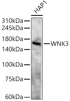
Western blot analysis of lysates from HAP1 cells, using WNK3 Rabbit pAb (CAB15507) at 1:500 dilution. Secondary antibody: HRP-conjugated Goat anti-Rabbit IgG (H+L) (CABS014) at 1:10000 dilution. Lysates/proteins: 25μg per lane. Blocking buffer: 3% nonfat dry milk in TBST. Detection: ECL Basic Kit (AbGn00020). Exposure time: 90s.
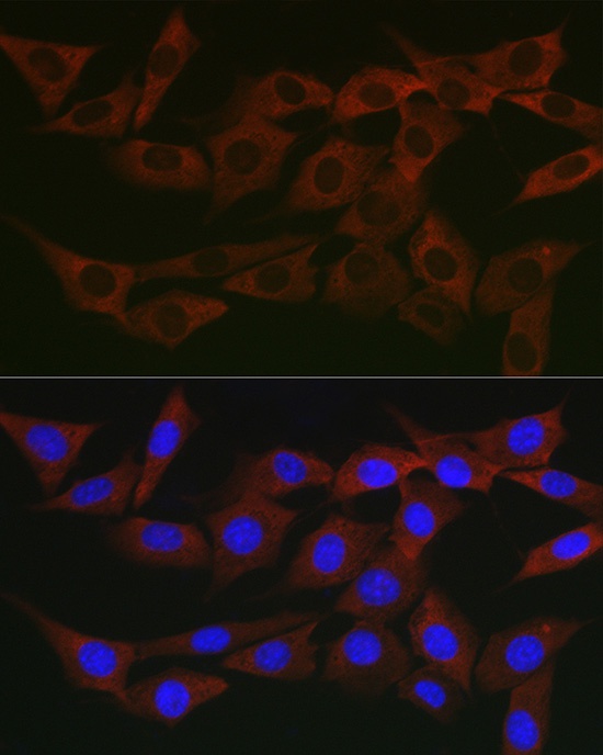
Immunofluorescence analysis of NIH/3T3 cells using WNK3 Rabbit pAb (CAB15507) at dilution of 1:200 (40x lens). Secondary antibody: Cy3-conjugated Goat anti-Rabbit IgG (H+L) (CABS007) at 1:500 dilution. Blue: DAPI for nuclear staining.
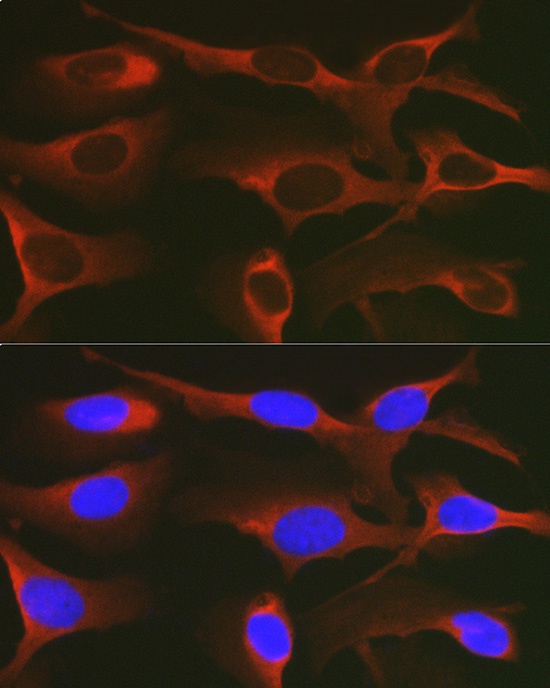
Immunofluorescence analysis of U2OS cells using WNK3 Rabbit pAb (CAB15507) at dilution of 1:200 (40x lens). Secondary antibody: Cy3-conjugated Goat anti-Rabbit IgG (H+L) (CABS007) at 1:500 dilution. Blue: DAPI for nuclear staining.