The YTHDF3 Antibody (CAB8395) is a high-quality antibody developed for reliable detection and analysis of target proteins. This antibody, produced in rabbits, shows high specificity and sensitivity towards human samples, making it an ideal choice for Western blot applications. By binding to the YTHDF3 protein, researchers can easily detect and analyze its expression in various cell types, providing valuable insights into RNA biology and potential therapeutic targets.YTHDF3 is a member of the YTH domain-containing family of proteins, which are important for mRNA metabolism and protein synthesis. It specifically recognizes and binds to m6A-modified mRNA, influencing its degradation or translation efficiency.
This antibody is validated for use in WB, IHC-P, IF/ICC, IP, ELISA applications and has demonstrated reactivity against Human, Mouse, Rat samples.
Product Name:
YTHDF3 Antibody
SKU:
CAB8395
Size:
20μL, 100μL
Reactivity:
Human, Mouse, Rat
Conjugate:
Unconjugated
Immunogen:
Recombinant protein (or fragment).This information is considered to be commercially sensitive.
This gene encodes a member of the YTH (YT521-B homology) domain protein family. The YTH domain is common in eukaryotes, is often found in the middle of the protein sequence, and may function in binding to RNA. Alternative splicing results in multiple transcript variants.
Purification Method
Affinity purification
Gene ID
253943
RRID
AB_2772933
Buffer Information
Store at -20℃. Avoid freeze / thaw cycles. Buffer: PBS containing 50% glycerol, preserved with proclin300 or sodium azide, pH 7.3.
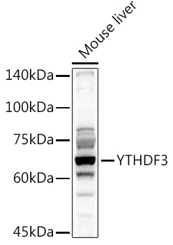
Western blot analysis of lysates from Mouse liver, using YTHDF3 Rabbit pAb (CAB8395) at 1:1000 dilution. Secondary antibody: HRP-conjugated Goat anti-Rabbit IgG (H+L) (CABS014) at 1:10000 dilution. Lysates/proteins: 25μg per lane. Blocking buffer: 3% nonfat dry milk in TBST. Detection: ECL Basic Kit (AbGn00020). Exposure time: 90s.
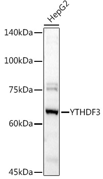
Western blot analysis of lysates from HepG2 cells, using YTHDF3 Rabbit pAb (CAB8395) at 1:1000 dilution. Secondary antibody: HRP-conjugated Goat anti-Rabbit IgG (H+L) (CABS014) at 1:10000 dilution. Lysates/proteins: 25μg per lane. Blocking buffer: 3% nonfat dry milk in TBST. Detection: ECL Basic Kit (AbGn00020). Exposure time: 90s.
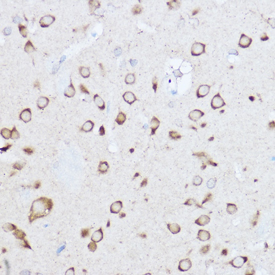
Immunohistochemistry analysis of paraffin-embedded Rat brain using YTHDF3 Rabbit pAb (CAB8395) at dilution of 1:500 (40x lens). High pressure antigen retrieval performed with 0.01M Citrate buffer (pH 6.0) prior to IHC staining.
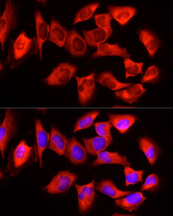
Immunofluorescence analysis of HeLa cells using YTHDF3 Rabbit pAb (CAB8395) at dilution of 1:50 (40x lens). Secondary antibody: Cy3-conjugated Goat anti-Rabbit IgG (H+L) (CABS007) at 1:500 dilution. Blue: DAPI for nuclear staining.
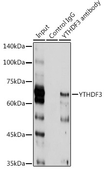
Immunoprecipitation analysis of 300 μg extracts of HT-1080 cells using 3 μg YTHDF3 antibody (CAB8395). Western blot was performed from the immunoprecipitate using YTHDF3 antibody (CAB8395) at a dilution of 1:1000.