Description
ADP/ATP Ratio Assay Kit (BA0127) (BA0127)
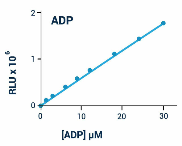
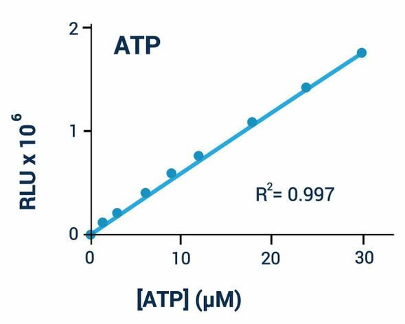
This ADP/ATP Ratio Assay Kit (SKU: BA0127) provides a rapid bioluminescent method for measuring ADP and ATP levels to screen for apoptosis, necrosis and cell proliferation in mammalian cells. Changes in the ADP/ATP ratio can be used to differentiate modes of cell death and viability, with proliferating cells showing increased ATP and decreased ADP and apoptotic or necrotic cells showing the reverse. The assay involves two steps: a working reagent first lyses the cells to release ATP and ADP, and in the presence of luciferase the ATP reacts with D-luciferin to produce light whose intensity is a direct measure of intracellular ATP. In the second step, the ADP is converted to ATP and then measured in the same way. This non-radioactive, homogeneous, cell-based assay is compatible with all culture media and liquid handling systems for high-throughput screening in 96-well and 384-well plates.
| Product Name: | ADP/ATP Ratio Assay Kit (BA0127) |
| SKU: | BA0127 |
| Detection Method: | Bioluminescent (luminometer) |
| Detection Range: | ADP/ATP ratio (relative luminescence) |
| Sample Type: | Mammalian cells (suspension or adherent) |
| Species Reactivity: | All |
| Assay Time: | Approximately 12 minutes (1 min ATP read, 10 min interval, then ADP read) |
| Kit Size: | 100 Assays |
| Equipment Required: | Microplate reader |
| Storage: | Store all reagents at -20°C. |
| Shelf Life: | 12 months after receipt. |
| Shipping: | Gel Pack |
A working reagent lyses cells to release ATP and ADP. Luciferase catalyses the reaction of ATP with D-luciferin to produce light proportional to ATP. In a second step, ADP is enzymatically converted to ATP and measured in the same way, allowing calculation of the ADP/ATP ratio.
- Safe, non-radioactive assay.
- Homogeneous and convenient 'mix-incubate-measure' assay with no wash or reagent transfer steps.
- Robust and amenable to high-throughput screening, with Z' factors of 0.5 and above routinely observed in 96-well and 384-well plates and readily automated for thousands of samples per day.
- Apoptosis and necrosis determination in cells.
- Cell proliferation: effects of cytokines, growth factors and nutrients.
- Drug discovery: high-throughput screening for anticancer drugs.
Note: The below protocol is a sample protocol. Protocols are specific to each batch/lot. For the correct instructions please follow the protocol included in your kit.
| Step | Procedure |
| 1 | Perform assays in a white opaque microplate for luminescent read-out and keep the time between luminescence measurements consistent across all samples. |
| 2 | Sample preparation: for suspension cells, transfer 10 µL of cultured cells (10^3-10^4) into a white opaque 96-well plate; for adherent cells, culture 10^3-10^4 cells in a white opaque microplate and remove the culture medium immediately before adding the ATP Reagent. |
| 3 | ATP assay: bring Assay Buffer, Substrate and Cosubstrate to room temperature and thaw the enzymes on ice or at 4°C; prepare ATP Reagent by mixing, per 96-well, 95 µL Assay Buffer with 1 µL Substrate, 1 µL Cosubstrate and 1 µL ATP Enzyme. |
| 4 | Add 90 µL ATP Reagent to each well, mix by tapping the plate and, after 1 minute, read the luminescence (RLU A) on a luminometer. |
| 5 | ADP assay: prepare ADP Reagent by mixing, per 96-well, 5 µL distilled water with 1 µL ADP Enzyme. |
| 6 | Ten minutes after reading RLU A, read the luminescence again (RLU B) to establish the residual ATP background. |
| 7 | Immediately after reading RLU B, add 5 µL ADP Reagent to each well, mix by tapping the plate or pipetting, and after 1 minute read the luminescence (RLU C). |
ADP/ATP Ratio = (RLU C - RLU B) / RLU A. Results interpretation guidelines: markedly elevated ATP with no significant increase in ADP indicates proliferation; lower ATP with increased ADP indicates apoptosis; and markedly lower ATP with greatly increased ADP indicates necrosis. Interpretation may vary with cell type and conditions.
| Component | Quantity | Storage |
| Assay Buffer | 10 mL | -20°C |
| Substrate | 120 µL | -20°C |
| Cosubstrate | 120 µL | -20°C |
| ATP Enzyme | 120 µL | -20°C |
| ADP Enzyme | 120 µL | -20°C |